. 2019 Oct;15(10):949-958.
doi: 10.1038/s41589-019-0342-2. Epub 2019 Aug 26.

CRISPR-Cas9 screens identify regulators of antibody-drug conjugate toxicity

Affiliations

CRISPR-Cas9 screens identify regulators of antibody-drug conjugate toxicity

C Kimberly Tsui et al. Nat Chem Biol. 2019 Oct.

Abstract

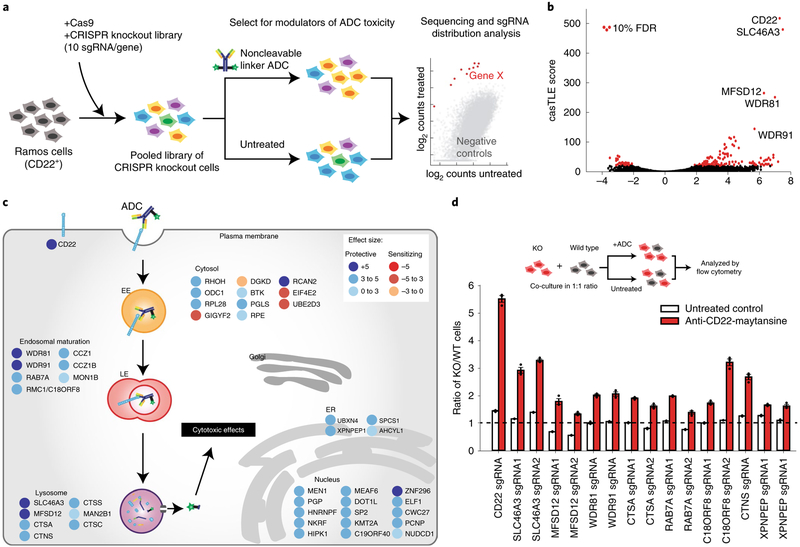
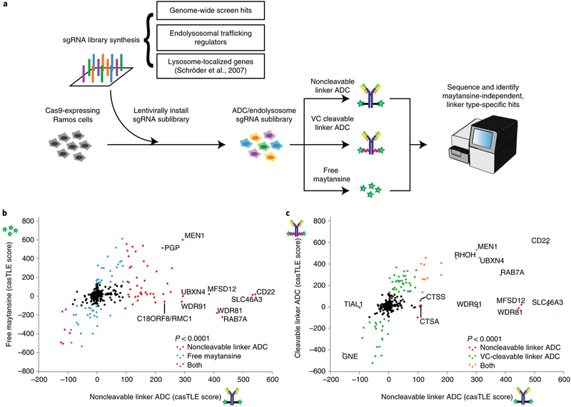
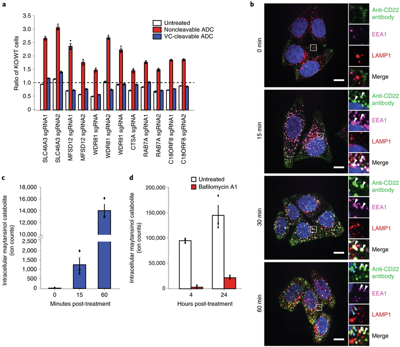
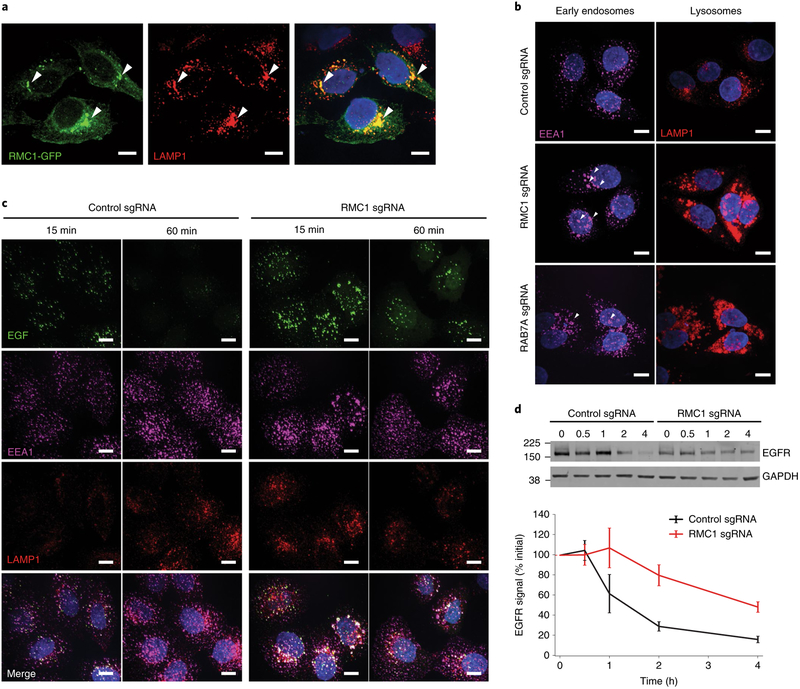
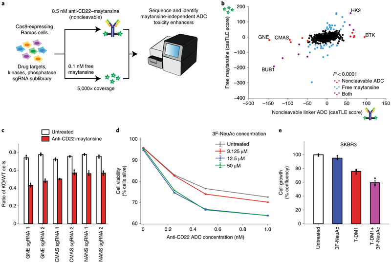
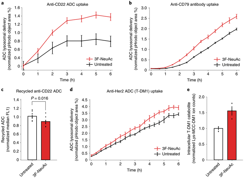
Antibody-drug conjugates (ADCs) selectively deliver chemotherapeutic agents to target cells and are important cancer therapeutics. However, the mechanisms by which ADCs are internalized and activated remain unclear. Using CRISPR-Cas9 screens, we uncover many known and novel endolysosomal regulators as modulators of ADC toxicity. We identify and characterize C18ORF8/RMC1 as a regulator of ADC toxicity through its role in endosomal maturation. Through comparative analysis of screens with ADCs bearing different linkers, we show that a subset of late endolysosomal regulators selectively influence toxicity of noncleavable linker ADCs. Surprisingly, we find cleavable valine-citrulline linkers can be processed rapidly after internalization without lysosomal delivery. Lastly, we show that sialic acid depletion enhances ADC lysosomal delivery and killing in diverse cancer cell types, including with FDA (US Food and Drug Administration)-approved trastuzumab emtansine (T-DM1) in Her2-positive breast cancer cells. Together, these results reveal new regulators of endolysosomal trafficking, provide important insights for ADC design and identify candidate combination therapy targets.


Conflict of interest statement

Competing interests

R.M.B. and D.R. are employees of Catalent Biologics. C.R.B. is a cofounder and Scientific Advisory Board member of Redwood Bioscience, which generated antibody-drug conjugates used in this work. Stanford University has filed a patent application based on the findings in this article.

Figures

Fig. 1 |
Fig. 1 |. Genome-wide CRISPR screen uncovers diverse endolysosomal regulators of ADC toxicity.
a, Schematic for the genome-wide screen. Ramos cells expressing Cas9 were infected with a lentiviral, genome-wide sgRNA library, the population was split and half the cells were treated with anti-CD22-maytansine ADC for three rounds, killing ~50% cells each round. The resulting populations were subjected to deep sequencing and analysis. The screen was performed in duplicate. b, Volcano plot of all genes indicating effect and confidence scores for genome-wide ADC screen. Effect and confidence scores determined by casTLE. c, Schematic for top 30 and selected endolysosomal trafficking regulator hits in genome-wide screen, color coded by effect size (calculated by casTLE). Subcellular localization is indicated according to gene ontology annotations. EE, early endosomes; LE, late endosomes; ER, endoplasmic reticulum. d, Validation of hits in Ramos cells using competitive-growth assays: cells expressing sgRNAs for knockout (KO) of indicated genes (mCherry+) and control (mCherry) were cocultured in 1:1 ratio. Cells were either treated with anti-CD22-maytansine ADC or left untreated for 3 d. Resulting ratio of KO:control was determined using flow cytometry. Data are presented as mean ± s.e.m. and are representative of two independent experiments performed in triplicate with consistent results. WT, wild type.
Fig. 2 |
Fig. 2 |. A subset of endolysosomal trafficking regulators are critical only for toxicity of the noncleavable linker ADC.
a, Schematic for sublibrary targeted screen using anti-CD22-maytansine (noncleavable), anti-CD22-VC-maytansine (cleavable) and free maytansine. The targeted sublibrary was designed, synthesized and lentivirally installed into Cas9-expressing Ramos cells. The Ramos cells were either treated with (1) anti-CD22-maytansine, (2) anti-CD22-VC-maytansine or (3) free maytansine or left untreated. The resulting populations were subjected to deep sequencing and analysis. The screens were performed in duplicate and at 1,000× coverage. b, Comparative analysis of results from anti-CD22-maytansine (noncleavable) and free maytansine sublibrary screens. Signed casTLE scores are reported. c, Comparative analysis of results from anti-CD22-maytansine (noncleavable) and anti-CD22-VC-maytansine (cleavable) sublibrary screens. Signed casTLE scores are reported.
Fig. 3 |
Fig. 3 |. Lysosomal delivery is not required for processing of VC cleavable linkers.
a, Validation of hits in Ramos cells using competitive-growth assays: cells expressing sgRNAs for KO of indicated genes (mCherry+) and control (mCherry) were cocultured in 1:1 ratio. Cells were either treated with anti-CD22-maytansine, anti-CD22-VC-maytansine (blue) or left untreated (white) for 3 d. Resulting ratio of KO:control was determined using flow cytometry. Data are presented as mean ± s.e.m. and are representative of two independent experiments performed in triplicate with consistent results. b, Internalization of anti-CD22 antibody conjugated to Alexa Fluor 488 (anti-CD22-AF488) was monitored by immunofluorescence microscopy in HeLa cells. Cells were incubated on ice with anti-CD22-AF488 for 20 min. Cells were then washed once with cold PBS and internalization was initiated by prewarmed media. Cells were washed and fixed at indicated time points after initiation of internalization. The bottom row is a magnified view of the white boxes, which shows the colocalization of the anti-CD22 antibodies with early endosomal marker, EEA1, and lysosomal marker, LAMP1. White arrows indicate colocalization of anti-CD22 antibody and EEA1; blue arrows indicate colocalization of anti-CD22 antibody and LAMP1. Scale bars, 10 μm. Images are representative of two independent experiments performed in triplicate. Quantification of colocalization is shown in Supplementary Fig. 3b. c, Maytansinol release from anti-CD22-VC-maytansine in CD22-expressing HeLa cells. Anti-CD22-VC-maytansine (10 nM) was added to cells and reactions were quenched at the indicated time points. The level of intracellular maytansinol catabolite was determined by LC-MS/MS; see Methods for detailed extraction and detection protocol. Maytansinol signal is normalized by cell number and internal standard MMAE. Data are presented as mean ± s.e.m. and are representative of three independent experiments performed in triplicate with consistent results. d, Maytansinol release from anti-CD22-VC-maytansine in CD22-expressing HeLa cells treated with bafilomycin A1. Cells were treated with 100 nM bafilomycin A1 or left untreated for 20 h. Cells were then incubated with anti-CD22-VC-maytansine for the indicated times. The level of intracellular maytansinol catabolite was determined by LC-MS/MS; see Methods for detailed extraction and detection protocol. Maytansinol signal is normalized by cell number and internal standard MMAE. Data are presented as mean ± s.e.m. and are representative of three independent experiments performed in triplicate with consistent results.
Fig. 4 |
Fig. 4 |. C18ORF8/RMC1 is required for endosomal trafficking and lysosomal delivery of ADCs.
a, C-terminal GFP-tagged RMC1 localizes to LAMP1-positive, late endosome/lysosomes, visualized by immunofluorescence and confocal microscopy. White arrows indicate sites of colocalization. The experiment was independently repeated twice, with similar results. Scale bars, 10 μm. b, CRISPRi-mediated knockdown of RMC1 or RAB7A in HeLa cells results in enlarged endosomes (EEA1) and lysosomes (LAMP1). The experiment was independently repeated twice, with similar results. Scale bars, 10 μm. c, Trafficking of EGF in RMC1-knockdown HeLa cells. Cells were serum-starved for 16 h, then were placed on ice and treated with EGF for 10 min. Cells were then washed once with cold PBS and internalization was initiated by prewarmed media. Cells were fixed at the indicated time and visualized by immunofluorescence and confocal microscopy. The experiment was independently repeated twice, with similar results. Scale bars, 10 μm. d, EGFR degradation in RMC1-knockdown HeLa cells. Cells were treated with EGF for 10 min, and then EGFR levels were examined at the indicated time points by western blot. EGFR levels were quantified with ImageJ software. Data are presented as mean ± s.e.m. and are representative of two independent experiments performed in triplicate with consistent results. See Supplementary Fig. 8 for uncropped western blot.
Fig. 5 |
Fig. 5 |. Inhibition of sialic acid synthesis sensitizes cells to CD22 ADC toxicity.
a, Schematic for targeted screen for identifying sensitizing hits. Ramos cells expressing Cas9 were infected with a lentiviral, sgRNA sublibrary targeting drug targets, kinases and phosphatases. Cells were then treated with either 0.5 nM anti-CD22-maytansine, 0.1 nM free maytansine or left untreated. The resulting populations were subjected to deep sequencing and analysis. The screen was performed in duplicate and at 5,000× coverage. b, Comparative analysis of results from anti-CD22 noncleavable linker ADC (0.5 nM) and free maytansine (0.1 nM) sublibrary screens performed at 5,000× coverage. c, Validation of hits in Ramos cells using competitive-growth assays. Cells expressing sgRNAs for KO of indicated genes (mCherry+) and control (mCherry) were cocultured in 1:1 ratio. Cells were either treated with anti-CD22 noncleavable ADC or left untreated for 3 d. Resulting ratio of KO:control was determined using flow cytometry. Data are presented as mean ± s.e.m. and are representative of two independent experiments performed in triplicate with consistent results. d, Cell viability of Ramos cells treated with noncleavable ADC and 3F-NeuAc at different concentrations. Cells were first treated with 3F-NeuAc at indicated concentrations for 48 h, then ADCs at indicated concentrations were added. Cell viability, determined by forward and side scatter gating for live cells, was assayed 72 h post-ADC addition by flow cytometry. Data are presented as mean ± s.e.m. and are representative of three independent experiments performed in triplicate with consistent results. e, Cell growth (normalized confluency percentage) of SKBR3 treated with 100 μM 3F-NeuAc, 2 nM T-DM1, or combination of both. Cells were pretreated with 100 μM 3F-NeuAc for 48 h, followed by 72-h incubation with 2 nM T-DM1. Confluency was determined by IncuCyte S3 Live cells analysis system and normalized to maximum confluency of the untreated condition at the end of the 5-d experiment. Data are presented as mean ± s.e.m. and are representative of two independent experiments performed in triplicate with consistent results.
Fig. 6 |
Fig. 6 |. Depletion of sialic acid enhances rate of ADC lysosomal delivery.
a, Internalization of pHrodo-labeled anti-CD22 ADC in wild-type Ramos cells pretreated with 12.5 μM 3F-NeuAc for 48 h (red) or untreated (black). pHrodo signal was measured using the IncuCyte S3 Live cells analysis system and normalized to cell area. Data are presented as mean ± s.e.m. and are representative of three independent experiments performed in triplicate with consistent results. b, Internalization of pHrodo-labeled anti-CD79 antibody in wild-type Ramos cells pretreated with 12.5 μM 3F-NeuAc for 48 h (red) or untreated (black). pHrodo signal was measured using the IncuCyte S3 Live cells analysis system and normalized to cell area. Data are presented as mean ± s.e.m. and are representative of two independent experiments performed in triplicate with consistent results. c, Recycling of anti-CD22 ADCs in Ramos cells pretreated with 12.5 μM 3F-NeuAc for 48 h (red) or untreated (white). Cells were treated with unlabeled anti-CD22 ADC for 1 h, acid washed to remove surface-bound ADCs and returned to 37 °C for 30 min to allow for ADC recycling. ADCs recycled back to the cell surface are detected by Alexa Fluor 488-labeled anti-human IgG antibodies and analyzed using flow cytometry. Data are presented as mean ± s.e.m. and are representative of two independent experiments with six replicates. d, Internalization of pHrodo-labeled T-DM1 in wild-type SKBR3 cells pretreated with 100 μM 3F-NeuAc for 48 h (red) or untreated (black). pHrodo signal was measured using the IncuCyte S3 Live cells analysis system and normalized to cell area. Data are presented as mean ± s.e.m. and are representative of two independent experiments performed in triplicate with consistent results. e, Accumulation of T-DM1 catabolite Lys-MCC-DM1 in wild-type SKBR3 cells treated with 100 μM 3F-NeuAc. SKBR3 cells were either pretreated with 100 μM 3F-NeuAc or left untreated for 48 h, followed by addition of 10 nM T-DM1. After 24 h, the level of intracellular T-DM1 catabolite, Lys-MCC-DM1, was determined by LC-MS/MS; see Methods for detailed extraction and detection protocol. Lyc-MCC-DM1 signal is normalized by cell number and the internal standard MMAE. Data are presented as mean ± s.e.m. and are representative of three independent experiments performed in triplicate with consistent results.

References

    1. Beck A, Goetsch L, Dumontet C & Corvaïa N Strategies and challenges for the next generation of antibody-drug conjugates. Nat. Rev. Drug Discov 16, 315–337 (2017). - PubMed
    1. Lyon R Drawing lessons from the clinical development of antibody-drug conjugates. Drug Discov. Today Technol 30, 105–109 (2018). - PubMed
    1. St Pierre CA, Leonard D, Corvera S, Kurt-Jones EA & Finberg RW Antibodies to cell surface proteins redirect intracellular trafficking pathways. Exp. Mol. Pathol 91, 723–732 (2011). - PMC - PubMed
    1. Ritchie M, Tchistiakova L & Scott N Implications of receptor-mediated endocytosis and intracellular trafficking dynamics in the development of antibody drug conjugates. MAbs 5, 13–21 (2013). - PMC - PubMed
    1. Loganzo F et al. Tumor cells chronically treated with a trastuzumabmaytansinoid antibody-drug conjugate develop varied resistance mechanisms but respond to alternate treatments. Mol. Cancer Ther 14, 952–963 (2015). - PubMed

Publication types

MeSH terms