Review
. 2019 Dec;224(9):3001-3018.
doi: 10.1007/s00429-019-01945-2. Epub 2019 Aug 26.

The cingulate cortex and limbic systems for emotion, action, and memory

Affiliations
Review

The cingulate cortex and limbic systems for emotion, action, and memory

Edmund T Rolls. Brain Struct Funct. 2019 Dec.

Abstract

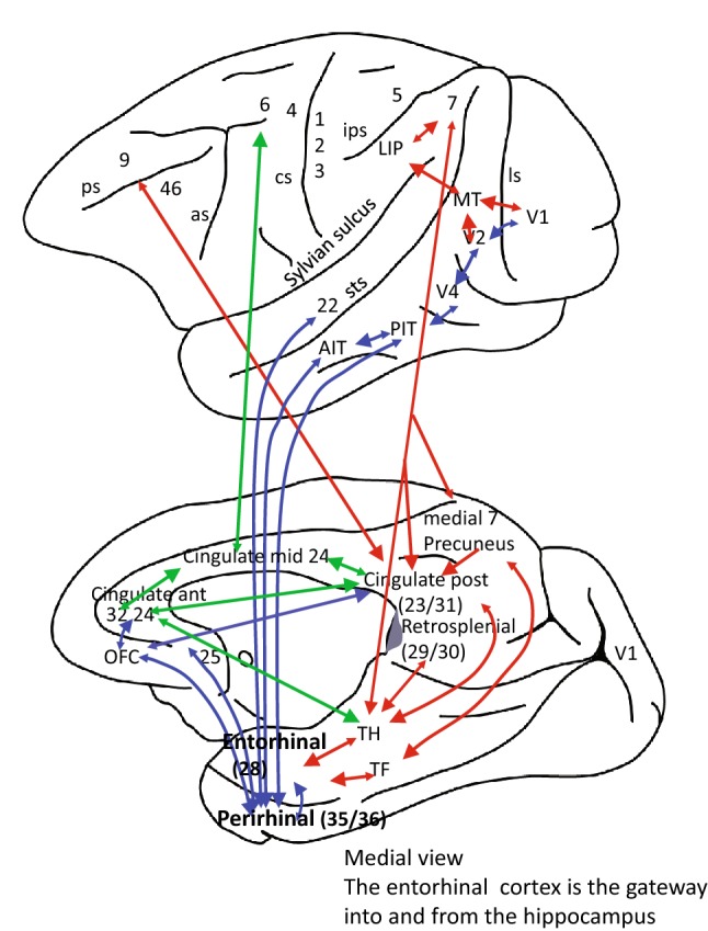
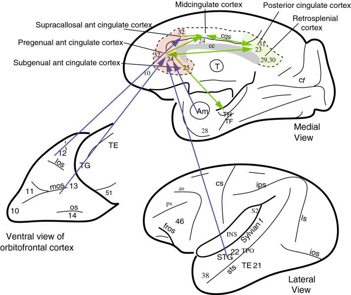
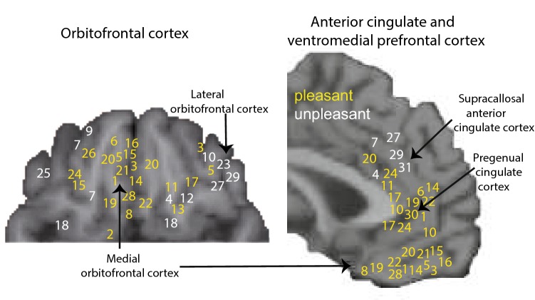
Evidence is provided for a new conceptualization of the connectivity and functions of the cingulate cortex in emotion, action, and memory. The anterior cingulate cortex receives information from the orbitofrontal cortex about reward and non-reward outcomes. The posterior cingulate cortex receives spatial and action-related information from parietal cortical areas. It is argued that these inputs allow the cingulate cortex to perform action-outcome learning, with outputs from the midcingulate motor area to premotor areas. In addition, because the anterior cingulate cortex connects rewards to actions, it is involved in emotion; and because the posterior cingulate cortex has outputs to the hippocampal system, it is involved in memory. These apparently multiple different functions of the cingulate cortex are related to the place of this proisocortical limbic region in brain connectivity.

Keywords: Cingulate cortex; Depression; Emotion; Hippocampus; Limbic systems; Memory; Orbitofrontal cortex.


Conflict of interest statement

The author declares no conflict of interest.

Figures

Fig. 1
Fig. 1
The connections of the anterior and posterior cingulate cortex with their input areas, and their outputs to the hippocampal memory system. A medial view of the macaque brain is shown below, and a lateral view is above. The green arrows show the convergence of reward or outcome information from the ACC and of information about actions from the posterior cingulate cortex to the midcingulate motor area, which then projects to premotor areas including the premotor cortex area 6 and the supplementary motor area. This provides connectivity for action–outcome learning. The anterior cingulate cortex receives reward outcome information from the orbitofrontal cortex (OFC). The posterior cingulate cortex (23 and 31) receives information about actions from the parietal cortex. This cingulate connectivity is compared with that of the hippocampus, which receives information from the ventral ‘what’ processing stream (blue) and the dorsal ‘where’ or ‘action’ processing stream (red), as described in the text. as, arcuate sulcus; cs, central sulcus; ips, intraparietal sulcus; ios, inferior occipital sulcus; ls, lunate sulcus; retrosplenial cortex (29, 30) is the small region in primates including humans behind the splenium of the corpus callosum shaded grey: it is present in rodents, which do not have a posterior cingulate cortex (Vogt 2009); sts, superior temporal sulcus; 4, 6, motor and premotor cortex. Developed from Rolls and Wirth (2018)
Fig. 2
Fig. 2
Connections of the anterior cingulate cortex shown on views of the primate brain (see text). The arrows show the main direction of connectivity, but there are connections in both directions. The supracallosal anterior cingulate cortex is also termed the anterior part of the midcingulate cortex, and is distinct from the posterior part of the midcingulate cortex (pMidcingulate). Connections reach the pregenual cingulate cortex especially from the medial/mid-orbitofrontal cortex; and reach the supracallosal anterior cingulate cortex especially from the lateral orbitofrontal cortex. Connections to the anterior cingulate cortex from the temporal lobe are from the (auditory) superior temporal gyrus (STG), from the visual and auditory cortex in the superior temporal sulcus; and from the amygdala. as, arcuate sulcus; cc, corpus callosum; cf., calcarine fissure; cgs, cingulate sulcus; cs, central sulcus; ls, lunate sulcus; ios, inferior occipital sulcus; mos, medial orbital sulcus; os, orbital sulcus; ps, principal sulcus; sts, superior temporal sulcus; Sf, Sylvian (or lateral) fissure (which has been opened to reveal the insula); Am, amygdala; INS, insula; TE (21), inferior temporal visual cortex; STG (22), superior temporal gyrus auditory association cortex; TF and TH, parahippocampal cortex; TPO, multimodal cortical area in the superior temporal sulcus; 38, TG, temporal pole cortex; 13, 11, medial orbitofrontal cortex; 12, lateral orbitofrontal cortex; 23, 31 posterior cingulate cortex; 29, 31 retrosplenial cortex; 51, olfactory (prepyriform and periamygdaloid) cortex. A cytoarchitectural map of the human cingulate cortex is provided in Fig. S1
Fig. 3
Fig. 3
Voxel-level parcellation of the left anterior cingulate cortex (ACC) based on its functional connectivity in healthy individuals with other brain areas. The correlations (r) are the distance from the centre of the circular plot. The pregenual and subcallosal subdivision (1, green) has strong functional connectivity with the medial orbitofrontal cortex and connected areas (AAL2 areas from OLF to OFCpost). The supracallosal subdivision (2, red) has strong functional connectivity with the lateral orbitofrontal cortex area IFGorb and with adjacent inferior frontal gyrus areas (IFGtriang to IFGoperc). The parcellation was similar on the right. The AAL2 is the automated anatomical labelling atlas, which shows the abbreviations used (Rolls et al. 2015)
Fig. 4
Fig. 4
Maps of subjective pleasure in the orbitofrontal cortex (ventral view) and anterior cingulate cortex (sagittal view). Yellow: sites where activations correlate with subjective pleasantness. White: sites where activations correlate with subjective unpleasantness. The numbers refer to effects found in specific studies. Taste: 1, 2; odour: 3–10; flavour: 11–16; oral texture: 17, 18; chocolate: 19; water: 20; wine: 21; oral temperature: 22, 23; somatosensory temperature: 24, 25; the sight of touch: 26, 27; facial attractiveness: 28, 29; erotic pictures: 30; laser-induced pain: 31. [After Grabenhorst and Rolls (2011) who provide references to the original studies.]

References

    1. Andersen RA. Coordinate transformations and motor planning in posterior parietal cortex. In: Gazzaniga MS, editor. The cognitive neurosciences. Cambridge: MIT Press; 1995. pp. 519–532.
    1. Andersen RA, Batista AP, Snyder LH, Buneo CA, Cohen YE. Programming to look and reach in the posterior parietal cortex. In: Gazzaniga MS, editor. The new cognitive neurosciences. Cambridge: MIT Press; 2000. pp. 515–524.
    1. Andrews-Hanna JR, Reidler JS, Sepulcre J, Poulin R, Buckner RL. Functional-anatomic fractionation of the brain’s default network. Neuron. 2010;65(4):550–562. doi: 10.1016/j.neuron.2010.02.005. - DOI - PMC - PubMed
    1. Auger SD, Maguire EA. Assessing the mechanism of response in the retrosplenial cortex of good and poor navigators. Cortex. 2013;49:2904–2913. doi: 10.1016/j.cortex.2013.08.002. - DOI - PMC - PubMed
    1. Balleine BW, Dickinson A. The role of incentive learning in instrumental outcome revaluation by sensory-specific satiety. Anim Learn Behav. 1998;26:46–59.