Review
. 2019 Dec;25(6):471-491.
doi: 10.1089/ten.TEB.2019.0182. Epub 2019 Oct 23.

Regenerative Therapies for Spinal Cord Injury

Affiliations
Review

Regenerative Therapies for Spinal Cord Injury

Nureddin Ashammakhi et al. Tissue Eng Part B Rev. 2019 Dec.

Abstract

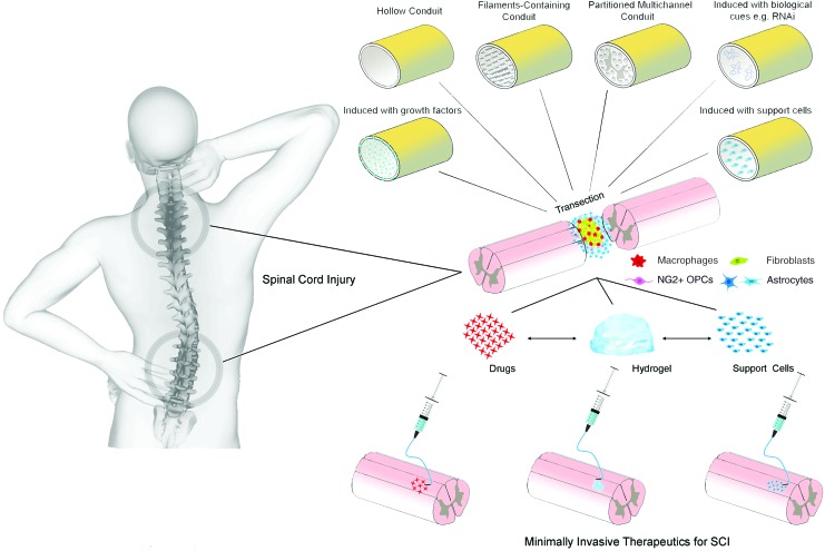
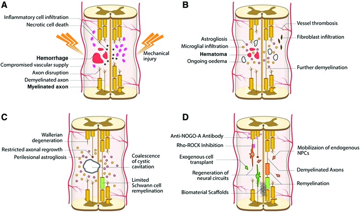
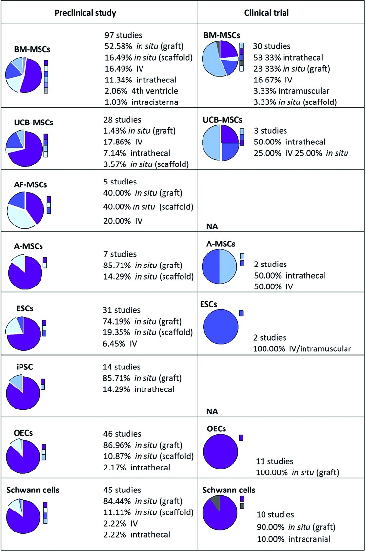
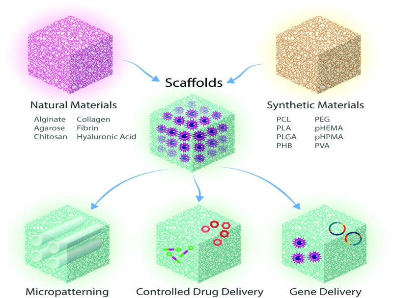
Spinal cord injury (SCI) is a serious problem that primarily affects younger and middle-aged adults at its onset. To date, no effective regenerative treatment has been developed. Over the last decade, researchers have made significant advances in stem cell technology, biomaterials, nanotechnology, and immune engineering, which may be applied as regenerative therapies for the spinal cord. Although the results of clinical trials using specific cell-based therapies have proven safe, their efficacy has not yet been demonstrated. The pathophysiology of SCI is multifaceted, complex and yet to be fully understood. Thus, combinatorial therapies that simultaneously leverage multiple approaches will likely be required to achieve satisfactory outcomes. Although combinations of biomaterials with pharmacologic agents or cells have been explored, few studies have combined these modalities in a systematic way. For most strategies, clinical translation will be facilitated by the use of minimally invasive therapies, which are the focus of this review. In addition, this review discusses previously explored therapies designed to promote neuroregeneration and neuroprotection after SCI, while highlighting present challenges and future directions. Impact Statement To date there are no effective treatments that can regenerate the spinal cord after injury. Although there have been significant preclinical advances in bioengineering and regenerative medicine over the last decade, these have not translated into effective clinical therapies for spinal cord injury. This review focuses on minimally invasive therapies, providing extensive background as well as updates on recent technological developments and current clinical trials. This review is a comprehensive resource for researchers working towards regenerative therapies for spinal cord injury that will help guide future innovation.

Keywords: biomaterials; cell therapy; minimally invasive; regeneration; spinal cord injury.


Conflict of interest statement

The authors acknowledge that they have no competing interests.

Figures

FIG. 1.
FIG. 1.
A schematic illustration showing different methods that can be used for the treatment of SCI. SCI, spinal cord injury. Adapted from Führmann et al. Color images are available online.
FIG. 2.
FIG. 2.
An illustration showing the development of pathophysiological changes following SCI. The acute phase takes 0–48 h and it involves hemorrhage, edema, and proapoptotic factors (A). This leads to further loss of function, more than that resulting from the initial insult occurs due to injury to neurons and oligodendrocytes. Astrocyte infiltration and release of additional proinflammatory factors are seen while demyelinated and injured axons begin to die back. In the late subacute (B) and intermediate (C) stages, microcystic cavities follow cell death. These cavities then coalesce forming barriers to regeneration in the chronic stage (>6 months). The final chronic stage scar, which is composed of a network of astrocytic processes and a dense fibrous deposit, acts as a physical and biochemical barrier to neurite outgrowth and cell migration. (D) A schematic illustration showing demyelination and axonal loss that follow SCI and various regenerative therapeutics that can be used including the use of biomaterials, cells, molecules, such as an anti-NOGO-A antibody treatment and Rho-ROCK inhibition, or agents to mobilize endogenous cells such as metformin. ROCK, Rho-associated protein kinase. Adapted from Ahuja et al. Color images are available online.
FIG. 3.
FIG. 3.
An illustration showing the global trends of clinical and preclinical studies in which cell transplantation is involved. Number of studies, type of cells (ESCs, BM-MSCs, A-MSCs, AF-MSCs, UCB-MSCs, or OECs), and routes of administration (IV, intrathecal, fourth ventricle, or intracisterna) are shown. When no data available, it was indicated as NA. AF-MSCs, amniotic fetal MSCs; A-MSCs, adipose tissue-derived mesenchymal stem cells; BM-MSCs, bone marrow-derived mesenchymal stem cells; ESCs, embryonic stem cells; IV, intravenous; OECs, olfactory ensheathing cells; UCB-MSCs, umbilical cord blood MSCs. Reproduced with permission from Vismara et al. Color images are available online.
FIG. 4.
FIG. 4.
Illustration showing that scaffolds (made of synthetic or natural biomaterials) can be used for the treatment of SCI, as micropatterned devices or as matrices combined with drug delivery, or gene delivery to enable guided tissue regeneration. Adapted from Iyer et al. Color images are available online.
FIG. 5.
FIG. 5.
Schematic illustration showing the main cellular targets of cell therapy in SCI and how this works (A), with particular focus on the mechanism by which stem cells act to achieve immunomodulatory action, differentiation into neurons and oligodendrocytes, or providing trophic factors that can support axonal regeneration (B). Reproduced with permission from Vismara et al. Color images are available online.
FIG. 6.
FIG. 6.
Schematic illustration showing bridge (A) or relay (B) formation during the repair process after the transplantation of cells for the treatment of SCI leading to neuronal connectivity. Reproduced with permission from Assinck et al. Color images are available online.
FIG. 7.
FIG. 7.
The use of magnetic rods in fibrin hydrogel to help guide regeneration that can be aligned upon exposure to magnetic field. (A). An illustration showing the method of preparation of injectable hybrid hydrogel. A unidirectional structure made of aligned rod-shaped, magnetoceptive microgels within the fibrin hydrogel is generated in situ by exposure to magnetic field. Following this, the surrounding liquid prehydrogel is crosslinked, so that microgel orientation is fixed to guide aligned cell ingrowth. Adapted from Rose et al. (B) Premixed fibroblasts can be seen to extend along the microgel axis (green), as visualized by stretched F-actin filaments (stained in red). Reproduced with permission Rose et al. Color images are available online.

References

    1. O'Shea T.M., Burda J.E., and Sofroniew M.V.. Cell biology of spinal cord injury and repair. J Clin Invest 127, 3259, 2017 - PMC - PubMed
    1. World Health Organization; The International Spinal Cord Society. International Perspectives on Spinal Cord Injury. Geneva, Switzerland: World Health Organization, 2013
    1. Cox C.S., Jr. Cellular therapy for traumatic neurological injury. Pediatr Res 83, 325, 2018 - PubMed
    1. Singh A., Tetreault L., Kalsi-Ryan S., Nouri A., and Fehlings M.G.. Global prevalence and incidence of traumatic spinal cord injury. Clin Epidemiol 6, 309, 2014 - PMC - PubMed
    1. Jazayeri S.B., Beygi S., Shokraneh F., Hagen E.M., and Rahimi-Movaghar V.. Incidence of traumatic spinal cord injury worldwide: a systematic review. Eur Spine J 24, 905, 2015 - PubMed

Publication types

Substances