

Synchronous Radial 1H and 23Na Dual-Nuclear MRI on a Clinical MRI System, Equipped With a Broadband Transmit Channel
- PMID: 31452649
- PMCID: PMC6710097
- DOI: 10.1002/cmr.b.21347
Synchronous Radial 1H and 23Na Dual-Nuclear MRI on a Clinical MRI System, Equipped With a Broadband Transmit Channel
Abstract
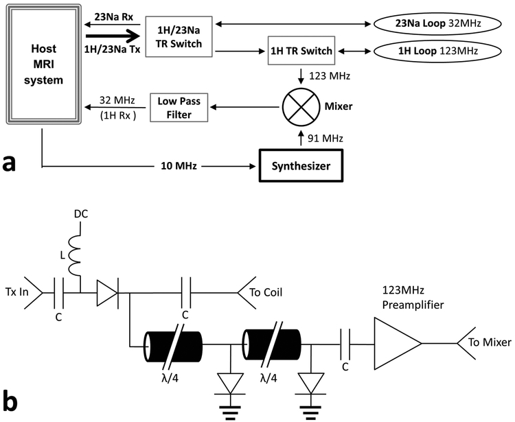
The purpose of this work was to synchronously acquire proton (1H) and sodium (23Na) image data on a 3T clinical MRI system within the same sequence, without internal modification of the clinical hardware, and to demonstrate synchronous acquisition with 1H/23Na-GRE imaging with Cartesian and radial k-space sampling. Synchronous dual-nuclear imaging was implemented by: mixing down the 1H signal so that both the 23Na and 1H signal were acquired at 23Na frequency by the conventional MRI system; interleaving 1H/23Na transmit pulses in both Cartesian and radial sequences; and using phase stabilization on the 1H signal to remove mixing effects. The synchronous 1H/23Na setup obtained images in half the time necessary to sequentially acquire the same 1H and 23Na images with the given setup and parameters. Dual-nuclear hardware and sequence modifications were used to acquire 23Na images within the same sequence as 1H images, without increases to the 1H acquisition time. This work demonstrates a viable technique to acquire 23Na image data without increasing 1H acquisition time using minor additional custom hardware, without requiring modification of a commercial scanner with multinuclear capability.
Keywords: dual-nuclear mri; metabolic imaging; multinuclear imaging; radiofrequency coils; sodium.
Figures


Similar articles
-
Theoretical basis for sodium and potassium MRI of the human heart at 1.5 T.Magn Reson Med. 1997 Oct;38(4):653-61. doi: 10.1002/mrm.1910380420. Magn Reson Med. 1997. PMID: 9324333
-
Hardware and Software Setup for Quantitative 23Na Magnetic Resonance Imaging at 3T: A Phantom Study.Sensors (Basel). 2024 Apr 24;24(9):2716. doi: 10.3390/s24092716. Sensors (Basel). 2024. PMID: 38732822 Free PMC article.
-
ECG-gated 23Na-MRI of the human heart using a 3D-radial projection technique with ultra-short echo times.MAGMA. 2004 May;16(6):297-302. doi: 10.1007/s10334-004-0038-8. Epub 2004 May 24. MAGMA. 2004. PMID: 15160295
-
High-performance radiofrequency coils for (23)Na MRI: brain and musculoskeletal applications.NMR Biomed. 2016 Feb;29(2):96-106. doi: 10.1002/nbm.3379. Epub 2015 Sep 24. NMR Biomed. 2016. PMID: 26404631 Free PMC article. Review.
-
23Na-MRI for Breast Cancer Diagnosis and Treatment Monitoring: A Scoping Review.Bioengineering (Basel). 2025 Feb 6;12(2):158. doi: 10.3390/bioengineering12020158. Bioengineering (Basel). 2025. PMID: 40001678 Free PMC article. Review.
Cited by
-
Repeatability of simultaneous 3D 1H MRF/23Na MRI in brain at 7 T.Sci Rep. 2022 Aug 19;12(1):14156. doi: 10.1038/s41598-022-18388-1. Sci Rep. 2022. PMID: 35986071 Free PMC article.
-
Simultaneous 3D acquisition of 1 H MRF and 23 Na MRI.Magn Reson Med. 2022 May;87(5):2299-2312. doi: 10.1002/mrm.29135. Epub 2021 Dec 31. Magn Reson Med. 2022. PMID: 34971454 Free PMC article.
-
Interleaved Whole Brain 23Na-MRI and 31P-MRSI Acquisitions at 7 Tesla.NMR Biomed. 2025 Mar;38(3):e70012. doi: 10.1002/nbm.70012. NMR Biomed. 2025. PMID: 39956139 Free PMC article.
-
Simultaneous proton magnetic resonance fingerprinting and sodium MRI.Magn Reson Med. 2020 Jun;83(6):2232-2242. doi: 10.1002/mrm.28073. Epub 2019 Nov 20. Magn Reson Med. 2020. PMID: 31746048 Free PMC article.
-
A Frequency Translation System for Multi-Channel, Multi-Nuclear MR Spectroscopy.IEEE Trans Biomed Eng. 2021 Jan;68(1):109-118. doi: 10.1109/TBME.2020.2997770. Epub 2020 Dec 21. IEEE Trans Biomed Eng. 2021. PMID: 32746012 Free PMC article.
References
-
- Rooney WD, Springer CS. 1991. The molecular environment of intracellular sodium: 23Na NMR relaxation. NMR in Biomedicine;4(5):227–245. - PubMed
-
- Weast RC, Astle MJ, Beyer WH. CRC Handbook of Chemistry and Physics: CRC press Boca Raton, FL; 1988.
-
- Ouwerkerk R, Bleich KB, Gillen JS, Pomper MG, Bottomley PA. 2003. Tissue sodium concentration in human brain tumors as measured with 23Na MR imaging. Radiology;227(2):529–37. - PubMed
-
- Ouwerkerk R, Jacobs MA, Macura KJ, Wolff AC, Stearns V, Mezban SD, Khouri NF, Bluemke DA, Bottomley PA. 2007. Elevated tissue sodium concentration in malignant breast lesions detected with non-invasive 23Na MRI. Breast Cancer Res Treat;106(2):151–60. - PubMed
-
- Borthakur A, Shapiro EM, Beers J, Kudchodkar S, Kneeland JB, Reddy R. 2000. Sensitivity of MRI to proteoglycan depletion in cartilage: comparison of sodium and proton MRI. Osteoarthr Cartilage;8(4):288–93. - PubMed
Grants and funding
LinkOut - more resources
Full Text Sources
Other Literature Sources