. 2019 Sep;18(3):2537-2547.
doi: 10.3892/ol.2019.10560. Epub 2019 Jul 4.

Detection and clinical significance of circulating tumor cells in patients with nasopharyngeal carcinoma

Affiliations

Detection and clinical significance of circulating tumor cells in patients with nasopharyngeal carcinoma

Zunbei Wen et al. Oncol Lett. 2019 Sep.

Abstract

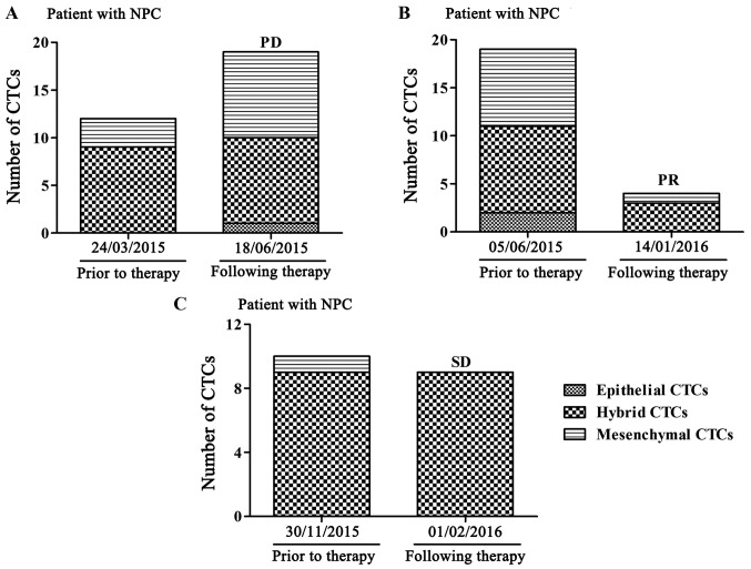
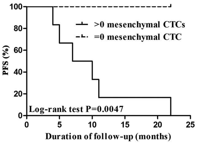
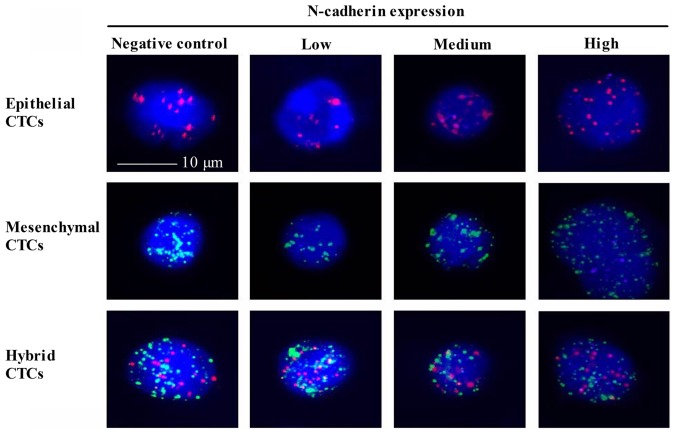
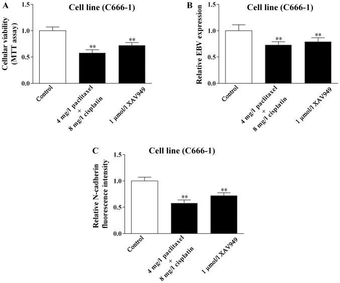
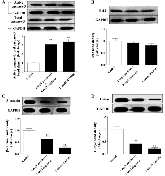
Nasopharyngeal carcinoma (NPC) is the most common cancer type originating in the nasopharynx, and varies notably from other cancer types of the head and neck in its occurrence, causes, clinical behavior and treatment. Significant effort has been made into understanding the biological properties of circulating tumor cells (CTCs), with previous studies demonstrating the critical role CTCs serve in the metastatic spread of carcinoma. However, associations between NPC and CTCs have not been completely elucidated. Therefore, in the present study, the CanPatrol™ CTC-enrichment technique and classical in situ hybridization assay were utilized to acquire, identify and classify CTCs from patients with NPC. Subsequently, the correlation between CTCs and the clinical indexes, progression-free survival (PFS), N-cadherin gene expression and the response to therapy were investigated. The present study then determined whether the Wnt/β-catenin signaling pathway served a role in therapy for NPC cells. Collectively, the research demonstrated that CTCs could be detected in patients with NPC. Additionally, CTCs exhibited a statistically significant association with the Epstein-Barr virus infection prior to therapy and Eastern Cooperative Oncology Group score following therapy. Furthermore, co-treatment with cisplatin and paclitaxel significantly decreased the number of CTCs. In addition, mesenchymal CTCs may serve as a predictor of PFS. Finally, the present study demonstrated that cisplatin combined with paclitaxel induced apoptosis and decreased the tumor markers in NPC cells through the Wnt/β-catenin signaling pathway. In conclusion, these data indicated that CTCs may serve as a biomarker in monitoring the therapeutic efficacy of treatments for NPC. Furthermore, the Wnt/β-catenin signaling pathway served a therapeutic role in the treatment of NPC.

Keywords: CanPatrol™ CTC-enrichment technique; N-cadherin; Wnt/β-catenin signaling pathway; circulating tumor cells; nasopharyngeal carcinoma.


Figures

Figure 1.
Figure 1.
Change in the number of CTCs in patients with NPC prior to and following treatment with cisplatin and paclitaxel. (A) CTCs were obtained from a patient with stage IVa NPC. (B) CTCs were obtained from a separate patient with stage IVa NPC. (C) CTCs were obtained from a patient with stage IVb NPC. CTCs, circulating tumor cells; NPC, nasopharyngeal carcinoma; PD, progressive disease; PR, partial response; SD, stable disease.
Figure 2.
Figure 2.
PFS according to CTC levels. CTCs/5 ml >0 vs. =0. CTC, circulating tumor cell; PFS, progression free survival; CTC, circulating tumor cells.
Figure 3.
Figure 3.
N-cadherin expression in different types of CTCs. N-cadherin (purple fluorescence) expression was depicted in different types of CTCs stained with CK18 (red fluorescence) and Twist (green fluorescence) biomarkers. The cells were analyzed using a ×100 oil objective. CTCs, circulating tumor cells.
Figure 4.
Figure 4.
Effect of paclitaxel combined with cisplatin on cellular viability, and EBV and N-cadherin expression on C666-1 cells. C666-1 cells were treated with 4 mg/l paclitaxel combined with 8 mg/l cisplatin or 1 µmol/l XAV949 for 48 h. (A) An MTT assay was applied to determine the change in the C666-1 cellular viability when treated with paclitaxel combined with cisplatin or XAV949. (B) An EBV-DNA assay was applied to determine the change in the C666-1 cellular EBV level when treated with paclitaxel combined with cisplatin or XAV949. (C) A Tri-color RNA-in situ hybridization assay was applied to determine the change in the C666-1 cellular N-cadherin level when treated with paclitaxel combined with cisplatin or XAV949. Data are presented as mean ± standard error of the mean of three independent experiments. One-way analysis of variance, followed by Dunnett's multiple-comparisons test, was utilized for analyzed differences among groups. **P<0.01 vs. control group. EBV, Epstein-Barr virus.
Figure 5.
Figure 5.
Effect of paclitaxel combined with cisplatin on apoptosis and Wnt/β-catenin signaling-associated proteins expression. C666-1 cells were incubated with indicated paclitaxel combined with cisplatin or XAV949 for 48 h. Immunoblots and bar graphs of indicated proteins levels of C666-1 cells. (A) Change in total caspase-3 and active caspase-3 expression following paclitaxel following paclitaxel combined with cisplatin or XAV949 treatment. (B) Change in BCL-2 expression following paclitaxel combined with cisplatin or XAV949 treatment. (C) Change in β-catenin expression following paclitaxel combined with cisplatin or XAV949 treatment. (D) Change in c-Myc expression following paclitaxel combined with cisplatin or XAV949 treatment. Data are presented as mean ± standard error of the mean of three independent experiments. One-way analysis of variance, followed by Dunnett's multiple-comparisons test, was utilized for analyzed differences among groups. **P<0.01 vs. control group. Bcl2, B-cell lymphoma.

References

    1. Cao SM, Simons MJ, Qian CN. The prevalence and prevention of nasopharyngeal carcinoma in China. Chin J Cancer. 2011;30:114–119. doi: 10.5732/cjc.010.10377. - DOI - PMC - PubMed
    1. Horikawa T, Yoshizaki T, Kondo S, Furukawa M, Kaizaki Y, Pagano JS. Epstein-Barr Virus latent membrane protein 1 induces Snail and epithelial-mesenchymal transition in metastatic nasopharyngeal carcinoma. Br J Cancer. 2011;104:1160–1167. doi: 10.1038/bjc.2011.38. - DOI - PMC - PubMed
    1. Chan AT. Nasopharyngeal carcinoma. Ann Oncol. 2010;21(Suppl 7):vii308–vii312. doi: 10.1093/annonc/mdq277. - DOI - PubMed
    1. Lin JH, Jiang CQ, Ho SY, Zhang WS, Mai ZM, Xu L, Lo CM, Lam TH. Smoking and nasopharyngeal carcinoma mortality: A cohort study of 101,823 adults in Guangzhou, China. BMC Cancer. 2015;15:906. doi: 10.1186/s12885-015-1902-9. - DOI - PMC - PubMed
    1. Sousa H, Mesquita L, Ribeiro J, Catarino R, Breda E, Medeiros R. Polymorphisms in host immune response associated genes and risk of nasopharyngeal carcinoma development in Portugal. Immunobiology. 2016;221:145–152. doi: 10.1016/j.imbio.2015.09.015. - DOI - PubMed