. 2019 Sep;18(3):2891-2896.
doi: 10.3892/ol.2019.10649. Epub 2019 Jul 22.

Matrine induces apoptosis in acute myeloid leukemia cells by inhibiting the PI3K/Akt/mTOR signaling pathway

Affiliations

Matrine induces apoptosis in acute myeloid leukemia cells by inhibiting the PI3K/Akt/mTOR signaling pathway

Yanmei Hao et al. Oncol Lett. 2019 Sep.

Abstract

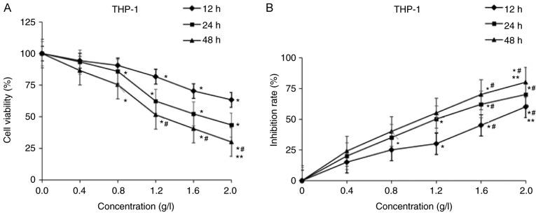
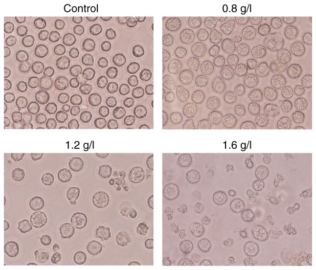
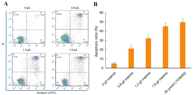
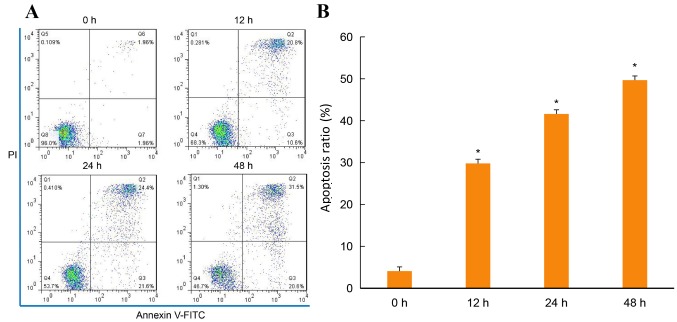
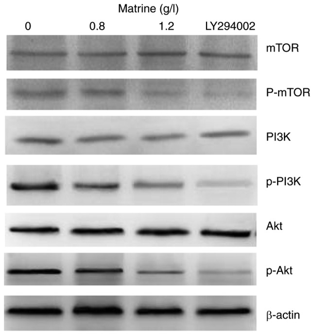
Matrine has been demonstrated to exert anticancer effects on acute myeloid leukemia (AML) cell lines. However, the mechanisms of matrine in AML remain largely unknown. The present study investigated the anticancer effects and underlying mechanisms of matrine on human AML cells in vitro. THP-1 cell lines were cultured and treated with different doses of matrine (0.4, 0.8, 1.2, 1.6 and 2.0 g/l). The effects of matrine on the cell proliferation were assessed by the Cell Counting Kit-8 assay. The apoptotic effects were evaluated by DAPI and annexin V/propidium iodide staining assays. The effects of the drug on phosphoinositide 3-kinase (PI3K)/protein kinase B (Akt)/ mechanistic target of rapamycin kinase (mTOR) protein expression were studied by western blot analysis. The results of the present study demonstrated that matrine suppressed the viability of THP-1 cells. The anticancer effects were identified to be dose-dependent and the IC50 value was 1.2 g/l in THP-1 cells. Matrine inhibited cell viability and induced cell apoptosis of AML cell lines in a dose- and time-dependent manner. In addition, it was observed that matrine decreased the expression of phosphorylated (p)-PI3K, p-Akt and p-mTOR in a concentration-dependent manner. However, the expression levels of PI3K, Akt and mTOR remained almost unaltered. These findings indicated that matrine may inhibit cell proliferation and induce apoptosis of AML cells and may be a novel effective chemotherapeutic agent against AML.

Keywords: acute myeloid leukemia; mammalian target of rapamycin; matrine; phosphoinositide 3-kinase; protein kinase B; signaling pathway.


Figures

Figure 1.
Figure 1.
Effects of matrine on the proliferation of acute myeloid leukemia cells. THP-1 cells were treated with 0, 0.4, 0.8, 1.2, 1.6 and 2.0 g/l matrine for 12, 24 and 48 h. Cell viability was assessed by a Cell Counting Kit-8 assay. (A) Cell viability rate. (B) Inhibition rate. Data are presented as the mean of at least three independent experiments. *P<0.05 vs. 0 g/l matrine; #P<0.05 vs. 0.4 g/l matrine; **P<0.05 vs. 1.2 g/l matrine.
Figure 2.
Figure 2.
Effects of matrine on cell morphology observed using phase contrast microscopy. Matrine induced changes in the morphology of THP-1 cells, such as the presence of autophagic vacuoles, which indicates that the drug exerts cytotoxic effects in these cells. Magnification, ×100.
Figure 3.
Figure 3.
Matrine induces apoptosis in a dose-dependent manner. Annexin V-FITC/PI staining was performed to assess apoptosis. Annexin V-negative/PI-negative (lower left quadrant), Annexin V-positive/PI-negative (lower right quadrant), Annexin V-positive/PI-positive (upper right quadrant) and Annexin V-positive/PI-positive (upper left quadrant) cells were considered as the viable, early-phase apoptotic, late-phase apoptotic and necrotic cells, respectively. The percentage of cells is presented in each quadrant. (A) THP-1 cells. (B) Representative histograms of apoptosis of THP-1 cells induced by matrine or LY294002 are presented. *P<0.05 vs. the control. The data are presented as the mean ± standard deviation of at least three independent experiments. FITC, fluorescein isothiocyanate; PI, propidium iodide.
Figure 4.
Figure 4.
Matrine induces apoptosis in a time-dependent manner. Annexin V-FITC/PI staining was performed to assess apoptosis. Annexin V-negative/PI-negative (lower left quadrant), Annexin V-positive/PI-negative (lower right quadrant), Annexin V-positive/PI-positive (upper right quadrant) and Annexin V-positive/PI-positive (upper left quadrant) cells were considered as the viable, early-phase apoptotic, late-phase apoptotic and necrosis cells, respectively. The percentage of cells is presented in each quadrant. (A) THP-1 cells. (B) Representative histograms of apoptosis of THP-1 cells induced by matrine are presented. *P<0.05 vs. 0 g/l matrine. The data are presented as the mean ± standard deviation of at least three independent experiments. FITC, fluorescein isothiocyanate; PI, propidium iodide.
Figure 5.
Figure 5.
Various concentrations of matrine affect the expression of PI3K/AKT pathway-associated proteins in THP-1 cells. Western blotting was performed in triplicate. Matrine decreased the expression levels of p-PI3K, p-Akt and p-mTOR in a concentration-dependent manner. However, no differences were revealed in the expression levels of PI3K, Akt and mTOR following treatment with matrine. PI3K, phosphoinositide 3-kinase; Akt, protein kinase B; mTOR, mechanistic target of rapamycin kinase; p, phosphorylated.

References

    1. Döhner H, Weisdorf DJ, Bloomfield CD. Acute myeloid leukemia. N Engl J Med. 2015;373:1136–1152. doi: 10.1056/NEJMra1406184. - DOI - PubMed
    1. Estey EH. Therapeutic options for acute myelogenous leukemia. Cancer. 2001;92:1059–1073. doi: 10.1002/1097-0142(20010901)92:5<1059::AID-CNCR1421>3.0.CO;2-K. - DOI - PubMed
    1. Jabo B, Morgan JW, Martinez ME, Ghamsary M, Wieduwilt MJ. Sociodemographic disparities in chemotherapy and hematopoietic cell transplantation utilization among adult acute lymphoblastic and acute myeloid leukemia patients. PLoS One. 2017;12:e0174760. doi: 10.1371/journal.pone.0174760. - DOI - PMC - PubMed
    1. Kwong YL, Au WY, Chim CS, Pang A, Suen C, Liang R. Arsenic trioxide- and idarubicin-induced remissions in relapsed acute promyelocytic leukemia: Clinicopathological and molecular features of a pilot study. Am J Hematol. 2001;66:274–279. doi: 10.1002/ajh.1057. - DOI - PubMed
    1. George B, Mathews V, Vishwabandhya A, Srivastava A, Chandy ML. Arsenic Trioxide (As2O3) in the treatment of patients with newly diagnosed acute promyelocytic leukemia (APML)-Toxicity and outcome. Blood. 2004;104:889. - PubMed