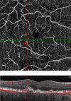
Vascularized drusen: a cross-sectional study
- PMID: 31452938
- PMCID: PMC6702713
- DOI: 10.1186/s40942-019-0187-6
Vascularized drusen: a cross-sectional study
Abstract
Background: To investigate whether neovascularization may arise and be detectable in drusen, as reported in histopathologic studies, by OCTA prior to developing exudation and to assess its prevalence in a cohort of patients with intermediate AMD.
Methods: Retrospective cross-sectional study of 128 patients with intermediate AMD recruited as part of a separate ongoing clinical trial conducted at multiple large tertiary referral retina clinics. One hundred and twenty-eight consecutive patients with exudative AMD in one eye and intermediate non-exudative AMD in the fellow eye were enrolled and analyzed between September 2015 and March 2017.
Results: SD-OCTA identified vascularization within drusen in 7 of 128 eyes, for a prevalence of 5.5%. A total of 12 instances of vascularized drusen were noted. Out of the 12 vascularized drusen noted, 7 were located in the parafoveal region or subfoveal region and 5 was in the extrafoveal region. 9 of 12 instances of vascularized drusen exhibited a uniform sub-RPE hyperreflectivity, whilst 3 of 12 exhibited more heterogenous reflectivity. In all 12 instances, FA images failed to identify the neovascular nature of vascularized drusen.
Conclusions: Our results demonstrate the utility of SD-OCTA for the diagnosis of vascularized drusen in patients with intermediate non-exudative AMD. Longitudinal studies are needed to delineate the evolution and conversion risk of these lesions over time, which can be of substantial clinical relevance.
Keywords: Age-related macular degeneration; Optical coherence tomography; Optical coherence tomography angiography; Retina; Retinal imaging; Vascularized drusen.
Conflict of interest statement
Competing interestsJSH: Financial Support: 4D Molecular Technologies, Acucela, Adverum, Aerie, Aerpio, Allegro, Apellis, Asclepix, Astellas, Bayer, BVI, Coda Therapeutix, Corcept, Daiichi Sankyo, Genentech/Roche, Genzyme, Heidelberg, Hemera, Janssen R&D, Kanghong, Kodiak, Neurotech, Notal Vision, Novartis, Ocular Therapeutix, Ophthotech, Optovue, Quark, Ra Pharmaceuticals, Regeneron, Regenxbio, Scifluor, Shire, Stealth Biotherapeutics, Thrombogenics, TLC, Tyrogenex. NW: Financial Support: Macula Vision Research Foundation, Topcon Medical Systems, Inc., Nidek Medical Products, Inc., Optovue, Inc. (Consultant), Carl Zeiss Meditec, Inc. JF: Optovue, Inc. (Patent, Personal Financial Interest and Consultant), Carl Zeiss Meditec, Inc. (Patent), Topcon Medical Systems, Inc. (Recipient). DBrown: Consultant – Genentech, Roche, Regeneron, Bayer, Novartis, Adverum, Santen, Samsung, Senju, Clearside Biomedical, Heidelberg, Optos, Zeiss, OHR, Regenxbio, ChengduKanghong Biotechnology, Apellis, Stealth Biotherapeutics; Grants/grants pending – Genentech, Roche, Regeneron, Clearside Biomedical, Heidelberg, Adverum, Novartis, OHR, Santen. DBoyer: Consulting fee – Genentech; Consulting fee or honorarium – OptoVue, Regeneron, Roche, Novartis, Alcon, Allergan, Bayer; Lecture fees – Allergan.
Figures


Similar articles
-
Age-related macular degeneration and risk factors for the development of choroidal neovascularisation in the fellow eye: a 3-year follow-up study.Ophthalmologica. 2011;226(3):110-8. doi: 10.1159/000329473. Epub 2011 Aug 3. Ophthalmologica. 2011. PMID: 21822000
-
Drusen Volume as a Predictor of Disease Progression in Patients With Late Age-Related Macular Degeneration in the Fellow Eye.Invest Ophthalmol Vis Sci. 2016 Apr;57(4):1839-46. doi: 10.1167/iovs.15-18572. Invest Ophthalmol Vis Sci. 2016. PMID: 27082298
-
Thinner choroid and greater drusen extent in retinal angiomatous proliferation than in typical exudative age-related macular degeneration.Am J Ophthalmol. 2013 Apr;155(4):743-9, 749.e1-2. doi: 10.1016/j.ajo.2012.11.001. Epub 2013 Jan 11. Am J Ophthalmol. 2013. PMID: 23317655
-
Exudative non-neovascular age-related macular degeneration.Graefes Arch Clin Exp Ophthalmol. 2021 May;259(5):1123-1134. doi: 10.1007/s00417-020-05021-y. Epub 2020 Nov 26. Graefes Arch Clin Exp Ophthalmol. 2021. PMID: 33242167
-
Optical Coherence Tomography Reflective Drusen Substructures Predict Progression to Geographic Atrophy in Age-related Macular Degeneration.Ophthalmology. 2016 Dec;123(12):2554-2570. doi: 10.1016/j.ophtha.2016.08.047. Epub 2016 Oct 25. Ophthalmology. 2016. PMID: 27793356 Free PMC article.
Cited by
-
Genomewide Association Study of Retinal Traits in the Amish Reveals Loci Influencing Drusen Development and Link to Age-Related Macular Degeneration.Invest Ophthalmol Vis Sci. 2022 Jul 8;63(8):17. doi: 10.1167/iovs.63.8.17. Invest Ophthalmol Vis Sci. 2022. PMID: 35857289 Free PMC article.
-
Multimodal Imaging of Subfoveal Pachydrusen Containing a Blood Flow Signal.Case Rep Ophthalmol Med. 2022 Jun 8;2022:5680913. doi: 10.1155/2022/5680913. eCollection 2022. Case Rep Ophthalmol Med. 2022. PMID: 35721663 Free PMC article.
References
LinkOut - more resources
Full Text Sources

