

Comparison of nonlinear microscopy and frozen section histology for imaging of Mohs surgical margins
- PMID: 31453008
- PMCID: PMC6701533
- DOI: 10.1364/BOE.10.004249
Comparison of nonlinear microscopy and frozen section histology for imaging of Mohs surgical margins
Abstract
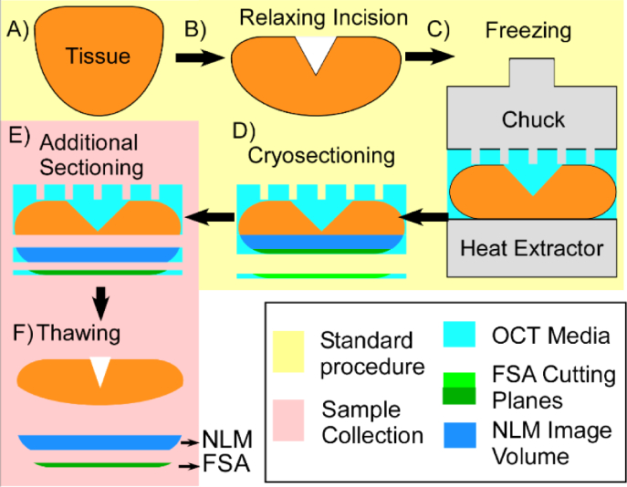
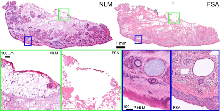
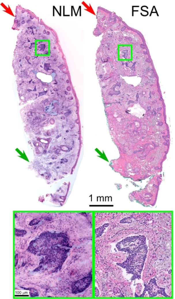
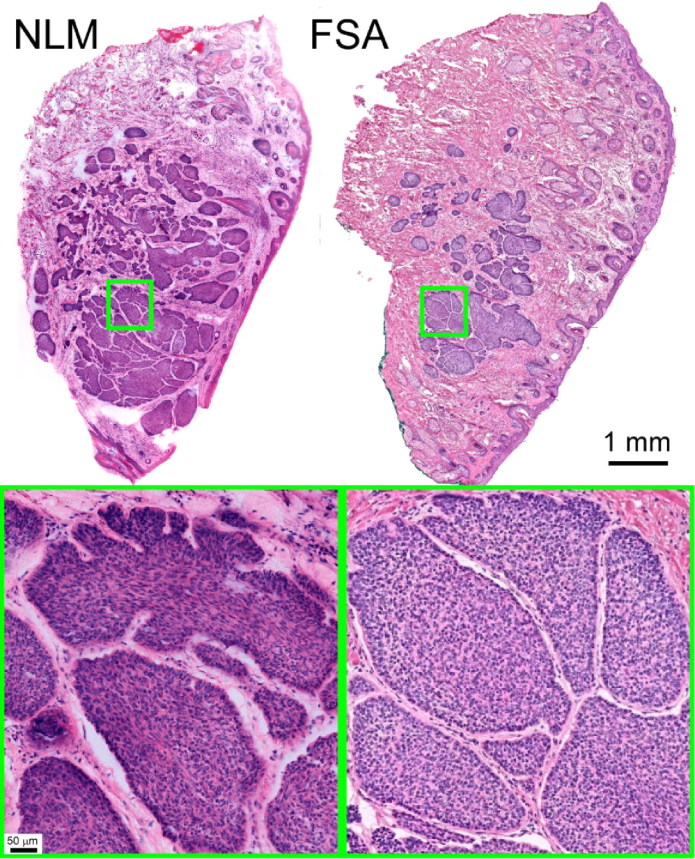
Mohs surgery uses en face frozen section analysis (FSA) with complete margin examination for the excision of select basal cell carcinomas (BCC), obtaining excellent cosmetic outcomes and extremely low recurrence rates. However, Mohs with FSA is time-consuming because of the need to iteratively perform cryosectioning on sequential excisions. Fluorescent microscopies can image tissue specimens without requiring physical sectioning, potentially reducing the time to perform Mohs surgery. We demonstrate a protocol for nonlinear microscopy (NLM) imaging of surgical specimens that combines dual agent staining, virtual H&E rendering, and video rate imaging. We also introduce a novel protocol that enables micron-level co-registration of NLM images with FSA histology, and demonstrate that NLM can reproduce similar features similar to FSA in BCC specimens with both negative and positive surgical margins. We show that the fluorescent labels can be extracted with conventional vacuum infiltration processing, enabling subsequent immunohistochemistry on fluorescently labeled tissue. This protocol can also be applied to evaluate the performance of NLM compared with FSA in a wide range of pathologies for intraoperative consultation.
Conflict of interest statement
The authors declare that there are no conflicts of interest related to this article.
Figures


References
-
- American Cancer Society, “Cancer Facts & Figures 2018,” http://www.cancer.org/research/cancerfactsstatistics/cancerfactsfigures2....
-
- Tierney E. P., Hanke C. W., “Cost effectiveness of Mohs micrographic surgery: review of the literature,” J. Drugs Dermatol. 8(10), 914–922 (2009). - PubMed
LinkOut - more resources
Full Text Sources
Other Literature Sources
Research Materials