. 2016 Jul 16;3(3):e48.
doi: 10.14440/jbm.2016.123. eCollection 2016.

Direct lentivirus injection for fast and efficient gene transfer into brown and beige adipose tissue

Affiliations

Direct lentivirus injection for fast and efficient gene transfer into brown and beige adipose tissue

Aileen Balkow et al. J Biol Methods. .

Abstract

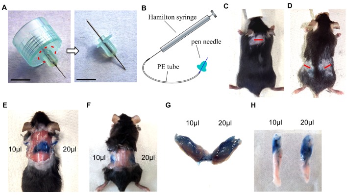
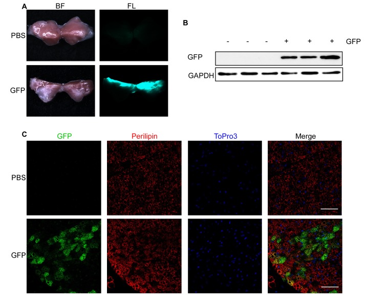
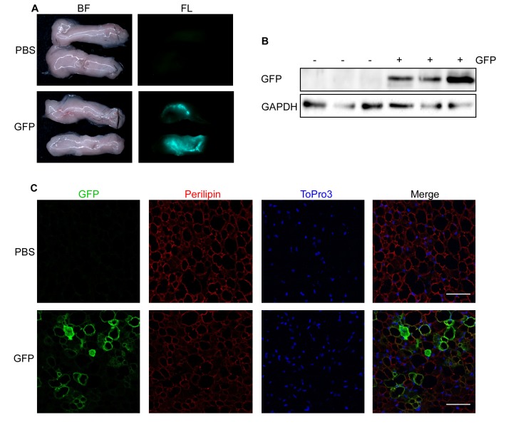
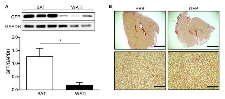
Brown adipose tissue is a special type of fat contributing to energy expenditure in human newborns and adults. Moreover, subcutaneous white adipose tissue has a high capacity to adapt an energy-consuming, brown-like/beige phenotype. Here, we developed an easy to handle and fast to accomplish method to efficiently transfer genes into brown and beige fat pads in vivo. Lentiviral vectors are directly injected into the target fat pad of anesthetized mice through a small incision using a modified, small needle connected to a microsyringe, which is well suited for infiltration of adipose tissues. Expression of the target gene can be detected in brown/beige fat one week after injection. The method can be applied within minutes to efficiently deliver transgenes into subcutaneous adipose tissues. Thus, this protocol allows for studying genes of interest in a timely manner in murine brown/beige fat and could potentially lead to new gene therapies for obesity.

Keywords: beige adipose tissue; brown adipose tissue; gene transfer; lentivirus.


Conflict of interest statement

The authors have declared no competing interests exist.

Figures

Figure 1
Figure 1
Set up of injection device and establishment of protocol. A. Original pen needle (dashed line indicates the cutting area) (left) and needle after cutting off plastic rim (right). Scale bar = 0.5 cm. B. Illustration showing setup of injection device. C and D. Pictures of anesthetized and shaved mouse, incision sites are marked by a red line in the interscapular region (C) or at the flanks, proximal of hips (D). E. Situs after injection of 10 μl and 20 μl Trypan blue solution in BAT. F. Situs after injection of 10 μl and 20 μl Trypan blue solution in the upper region of WATi. G. Dissected BAT from (E). H. Dissected WATi from (F).
Figure 2
Figure 2
GFP fluorescence in interscapular BAT after lentivirus injection. A-C. 1000 ng RT of lentivirus carrying GFP under control of a CMV promoter (GFP) in 25 μl HBSS or 25 μl PBS were injected into each interscapular BAT lobe of 4-week-old male mice and analyzed after 1 week. A. Bright field (BF, left) and fluorescent images (FL, right) of PBS (upper panel) and GFP (lower panel) injected BAT. B. GFP expression assessed by Western blotting. Respective blots of GFP and the loading control GAPDH are shown. C. GFP expression assessed by immunohistological staining. PFA-fixed BAT sections were triple stained with antibodies directed against GFP (green) and Perilipin (red) as well as with ToPro3 (blue) to stain the nuclei. Scale bar = 50 µm.
Figure 3
Figure 3
Expression of GFP in inguinal WAT (WATi) after lentivirus injection. A-C. 1000 ng RT of lentivirus carrying GFP under control of a CMV promoter (GFP) in 30 μl HBSS or 30 μl PBS were injected into each WATi pad of 10-week-old male mice and analyzed after 1 week. A. Bright field (BF, left) and fluorescent images (FL, right) of PBS (upper panel) and GFP (lower panel) injected WATi. B. GFP expression assessed by Western blotting. Respective blots of GFP and the loading control GAPDH are shown. C. GFP expression assessed by immunohistological staining. PFA-fixed WATi sections were triple stained with antibodies directed against GFP (green) and Perilipin (red) as well as with ToPro3 (blue) to stain the nuclei. Scale bar = 50 µm.
Figure 4
Figure 4
Comparison of GFP expression in BAT and WATi and Sirius Red staining of BAT. A. GFP expression in BAT and WATi after injection of 1000 ng RT of lentivirus carrying GFP into each fat lobe assessed by Western blotting (top) and quantitative analysis (bottom). * P < 0.05 (n = 3). B. Representative BAT sections of PBS- and GFP-injected mice, stained with Sirius Red for collagen (I and III) fibres. Scale bar top = 1 mm; scale bar bottom = 100 µm.

References

    1. Ng M., Fleming T., Robinson M., Thomson B., Graetz N., et al. Global, regional, and national prevalence of overweight and obesity in children and adults during 1980-2013: a systematic analysis for the Global Burden of Disease Study 2013. Lancet. 2014;384:766–781. doi: 10.1016/S0140-6736(14)60460-8. PMID: 24880830. - DOI - PMC - PubMed
    1. Pfeifer A., Hoffmann L. S. Brown, beige, and white: the new color code of fat and its pharmacological implications. Annu Rev Pharmacol Toxicol. 2014;55:207–227. doi: 10.1146/annurev-pharmtox-010814-124346. PMID: 25149919. - DOI - PubMed
    1. Cannon B., Nedergaard J. Brown adipose tissue: function and physiological significance. Physiol Rev. 2004;84:277–359. doi: 10.1152/physrev.00015.2003. PMID: 14715917. - DOI - PubMed
    1. Klingenspor M., Herzig S., Pfeifer A. Brown fat develops a brite future. Obes Facts. 2012;5:890–896. doi: 10.1159/000346337. PMID: 23296106. - DOI - PubMed
    1. van Marken Lichtenbelt D., (Wouter), Vanhommerig J. W., Smulders N. M., Drossaerts J. M. A. F. L., Kemerink G. J., et al. Cold-activated brown adipose tissue in healthy men. N Engl J Med. 2009;360:1500–1508. doi: 10.1056/NEJMoa0808718. PMID: 19357405. - DOI - PubMed

LinkOut - more resources