. 2017 Nov 3;4(4):e81.
doi: 10.14440/jbm.2017.206. eCollection 2017.

IL-6-HaloTag® enables live-cell plasma membrane staining, flow cytometry, functional expression, and de-orphaning of recombinant odorant receptors

Affiliations

IL-6-HaloTag® enables live-cell plasma membrane staining, flow cytometry, functional expression, and de-orphaning of recombinant odorant receptors

Franziska Noe et al. J Biol Methods. .

Abstract

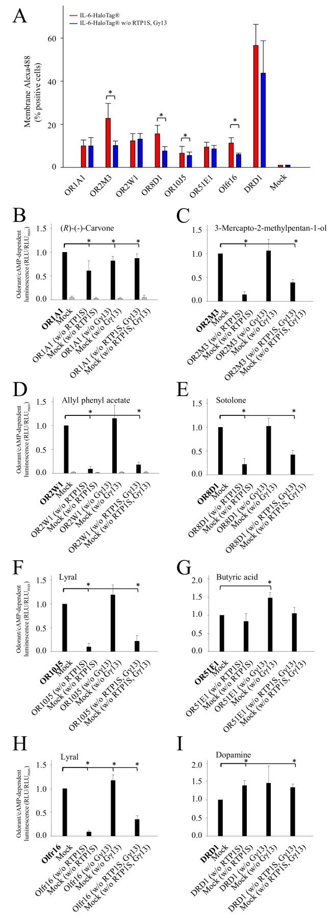
The assignment of cognate odorant/agonist pairs is a prerequisite for an understanding of odorant coding at the receptor level. However, the identification of new ligands for odorant receptors (ORs) in cell-based assays has been challenging, due to their individual and rather sub-optimal plasma membrane expression, as compared with other G protein-coupled receptors. Accessory proteins, such as the chaperone RTP1S, or Ric8b, have improved the surface expression of at least a portion of ORs. Typically, recombinant ORs carry N-terminal tags, which proved helpful for their functional membrane expression. The most common tag is the 'Rho-tag', representing an N-terminal part of rhodopsin, but also 'Lucy-' or 'Flag-tag' extensions have been described. Here, we used a bi-functional N-terminal tag, called 'interleukin 6 (IL-6)-HaloTag®', with IL-6 facilitating functional cell surface expression of recombinant ORs, and the HaloTag® protein, serving as a highly specific acceptor for cell-impermeant or cell-permeant, fluorophore-coupled ligands, which enable the quantification of odorant receptor expression by live-cell flow cytometry. Our experiments revealed on average an about four-fold increased surface expression, a four-fold higher signaling amplitude, and a significantly higher potency of odorant-induced cAMP signaling of six different human IL-6-HaloTag®-ORs across five different receptor families in NxG 108CC15 cells, as compared to their Rho-tag-HaloTag® constructs. We observed similar results in HEK-293 cells. Moreover, screening an IL-6-HaloTag®-odorant receptor library with allyl phenyl acetate, revealed both known receptors as best responders for this compound. In summary, the IL-6-HaloTag® represents a promising tool for the de-orphaning of ORs.

Keywords: N-terminal epitope tags; Rho-tag; cAMP assay; flow cytometry.


Conflict of interest statement

Competing interests: The authors have declared that no competing interests exist.

Figures

Figure 1.
Figure 1.. Concentration-response relations for selected odorant/OR combinations with different N-terminal tags in NxG 108CC15 cells.
A. Schemes of receptor constructs, carrying either IL-6-HaloTag® or Rho-tag-HaloTag®. B. OR1A1 with (R)-(-)-carvone. C. OR2M3 with 3-mercapto-2-methylpentan-1-ol. D. OR2W1 with allyl phenyl acetate. E. OR8D1 with sotolone. F. OR10J5 with lyral. G. OR51E1 with butyric acid. H. Olfr16 with lyral [29]. I. DRD1 with dopamine. EC50 values (Table 1) were derived from fitting a function to concentration-response data by means of non-linear regression. Shown are mean ± SD (n = 3–5). Data were normalized to each receptor’s maximum amplitude with the N-terminal tag IL-6-HaloTag®. Red colored curves indicate receptors with the N-terminal tag IL-6-HaloTag®, blue with Rho-tag-HaloTag®, and black curves indicate the empty plasmid control (Mock).
Figure 2.
Figure 2.. Normalized efficacies of receptors carrying the epitope tags IL-6-HaloTag® or Rho-tag-HaloTag® in HEK-293 cells.
A. Shown are the normalized amplitudes of receptors carrying either the IL-6-HaloTag® (red bars) or the Rho-tag-HaloTag® (blue bars), when challenged with their reported agonist: OR1A1 with (R)-(-)-carvone (1000 μM); OR2M3 with 3-mercapto-2-methylpentan-1-ol (60 μM); OR2W1 with allyl phenyl acetate (300 μM); OR8D1 with sotolone (300 μM); OR10J5 with lyral (300 μM); OR51E1 with butyric acid (300 μM); Olfr16 with lyral (300 μM); and DRD1 with dopamine (10 nM). Shown are means ± SD (n = 3). Data were normalized to each receptor’s maximum amplitude with the N-terminal tag IL-6-HaloTag®. Mock, empty plasmid control (black bars). B. Shown are differences in odorant responses averaged over all tested ORs carrying either the IL-6-HaloTag® (red bar) or the Rho-tag-HaloTag® (blue bar).
Figure 3.
Figure 3.. Analysis of the cell surface expression of investigated ORs with both N-terminal tags.
A. Flow cytometry analysis of transiently transfected NxG 108CC15 cells showing a detectable fluorescence of membrane-impermeable HaloTag® Ligand-Alexa488, suggesting different cell surface expression of ORs carrying different N-terminal tags. Data are means ± SD (n = 3–6). Mock, empty plasmid control. *P < 0.05 as compared to Rho-tag-HaloTag®. B. Differences averaged over all tested ORs, carrying either the IL-6-HaloTag® (red bar), or the Rho-tag-HaloTag® (blue bar), and normalized to the Rho-tag-HaloTag® ORs. *P < 0.05 as compared to Rho-tag-HaloTag®. C. Fluorescence confocal image of NxG 108CC15 cells expressing IL-6-HaloTag®-OR8D1, labeled with membrane-impermeable HaloTag® Ligand-Alexa488, and treated with endocytosis blocker Dynasore. D. Fluorescence confocal image of NxG 108CC15 cells expressing IL-6-HaloTag®-OR8D1, labeled with membrane-impermeable HaloTag® Ligand-Alexa488, but without endocytosis blocker Dynasore.
Figure 4.
Figure 4.. The influence of accessory proteins on the OR cAMP signaling in NxG 108CC15 cells.
Flow cytometry analysis of the cell surface expression of investigated ORs (A). The data represent the percentage of NxG 108CC15 transiently transfected cells with a detectable membrane-impermeable HaloTag® Ligand-Alexa488-dependent fluorescence, suggesting cell surface expression of OR, as determined by flow cytometry. Red bars indicate the transfection with all accessory proteins and blue bars indicate the transfection without (w/o) RTP1S and Gγ13. Shown are mean ± SD (n = 3–6). Mock, empty plasmid control. cAMP-luminescence measurements of OR1A1 with (R)-(-)-carvone (1000 μM) (B), OR2M3 with 3-mercapto-2-methylpentan-1-ol (60 μM) (C), OR2W1 with allyl phenyl acetate (300 μM) (D), OR8D1 with sotolone (300 μM) (E), OR10J5 with lyral (300 μM) (F), OR51E1 with butyric acid (300 μM) (G), Olfr16 with lyral (300 μM) (H), and DRD1 with dopamine (10 nM) (I). All receptors were tagged with IL-6-HaloTag®. Data were normalized to each receptor with co-transfection of RTP1S and Gγ13. Shown are mean ± SD (n = 3). Black bars are receptor responses in the presence of RTP1S and Gγ13, and grey bars indicate empty plasmid control (Mock). *P < 0.05 as compared to receptor responses in the presence of RTP1S and Gγ13.
Figure 5.
Figure 5.. Allyl phenyl acetate at 100 μM activates two receptors out of 579 odorant receptor variants.
Screening of allyl phenyl acetate (100 μM) against 579 OR variants in NxG 108CC15 cells. Data (n = 1 in duplicates) were normalized to the maximum responding OR (OR1A1). OR families are color-coded and sorted in ascending numerical order. Dashed lines indicate 2- and 3σ-thresholds. Mock, empty plasmid control.

References

    1. Buck L, Axel R. (1991) A novel multigene family may encode odorant receptors: a molecular basis for odor recognition. Cell 65: 175-187. doi: 10.1016/0092-8674(91)90418-X. PMID: - DOI - PubMed
    1. Lu M, Staszewski L, Echeverri F, Xu H, Moyer BD. (2004) Endoplasmic reticulum degradation impedes olfactory G-protein coupled receptor functional expression. BMC Cell Biol 5: 34. doi: 10.1186/1471-2121-5-34. PMID: - DOI - PMC - PubMed
    1. McClintock TS, Sammeta N. (2003) Trafficking prerogatives of olfactory receptors. Neuroreport 14: 1547-1552. doi: 10.1097/01.wnr.0000085904.20980.e1. PMID: - DOI - PubMed
    1. Keller A, Zhuang H, Chi Q, Vosshall LB, Matsunami H. (2007) Genetic variation in a human odorant receptor alters odour perception. Nature 449: 468-472. doi: 10.1038/nature06162. PMID: - DOI - PubMed
    1. Jaeger SR, McRae JF, Bava CM, Beresford MK, Hunter D, et al. (2013) A Mendelian trait for olfactory sensitivity affects odor experience and food selection. Curr Biol 23: 1601-1605. doi: 10.1016/j.cub.2013.07.030. PMID: - DOI - PubMed

LinkOut - more resources