. 2020 Feb;68(2):246-262.
doi: 10.1002/glia.23714. Epub 2019 Aug 27.

Neuron-generated thrombin induces a protective astrocyte response via protease activated receptors

Affiliations

Neuron-generated thrombin induces a protective astrocyte response via protease activated receptors

Padmesh S Rajput et al. Glia. 2020 Feb.

Abstract

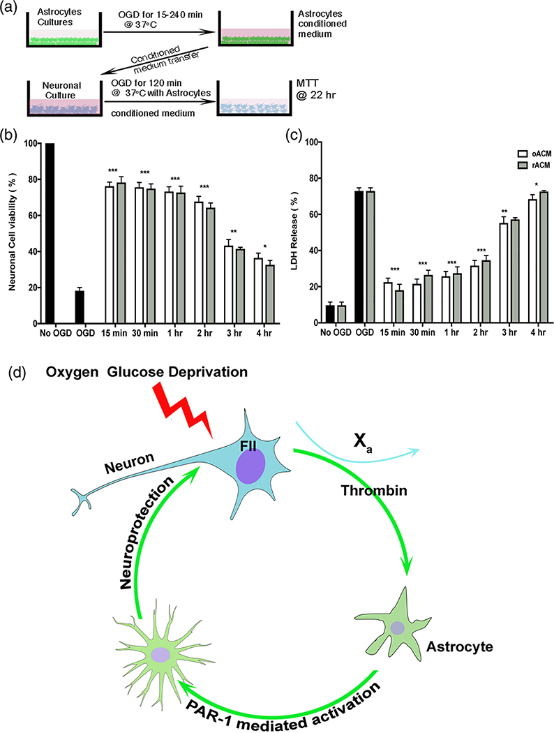
Astrocytes protect neurons during cerebral injury through several postulated mechanisms. Recent therapeutic attention has focused on enhancing or augmenting the neuroprotective actions of astrocytes but in some instances astrocytes can assume a neurotoxic phenotype. The signaling mechanisms that drive astrocytes toward a protective versus toxic phenotype are not fully known but cell-cell signaling via proteases acting on cell-specific receptors underlies critical mechanistic steps in neurodevelopment and disease. The protease activated receptor (PAR), resides in multiple brain cell types, and most PARs are found on astrocytes. We asked whether neuron-generated thrombin constituted an important astrocyte activation signal because our previous studies have shown that neurons contain prothrombin gene and transcribed protein. We used neuron and astrocyte mono-cell cultures exposed to oxygen-glucose deprivation and a model of middle cerebral artery occlusion. We found that ischemic neurons secrete thrombin into culture media, which leads to astrocyte activation; such astrocyte activation can be reproduced with low doses of thrombin. Media from prothrombin-deficient neurons failed to activate astrocytes and adding thrombin to such media restored activation. Astrocytes lacking PAR1 did not respond to neuron-generated thrombin. Induced astrocyte activation was antagonized dose-dependently with thrombin inhibitors or PAR1 antagonists. Ischemia-induced astrocyte activation in vivo was inhibited after neuronal prothrombin knockout, resulting in larger strokes. Restoring prothrombin to neurons with a lentiviral gene vector restored astrocyte activation and reduced stroke damage. We conclude that neuron-generated thrombin, released during ischemia, acts via PAR1 and may cause astrocyte activation and paracrine neuroprotection.

Keywords: astrocyte activation; ischemia; neuroprotection; thrombin.


Figures

FIGURE 1
FIGURE 1
Neuron-generated thrombin is critical for astrocyte mediated neuroprotection. (a) in NestinCreERT2FII f/f mice (Figure S1) we induced prothrombin knockout with tamoxifen (or vehicle, randomized) for 5 days. At random, 9 days later the animals received stereotactic injection of lentivirus containing the FII gene, or an empty vector, or no viral injections. All animals underwent MCAo 21 days after tamoxifen or vehicle and sacrifice 24 hr after MCAo. (b) Mid-parietal sections stained for GFAP (red) and lipocalin-2 (LCN2) (green) show robust GFAP expression in the ischemic territory of vehicle-treated NestinCreERT2FIIf/f mice, but less so after tamoxifen-induced FII KO. In the ischemic zone, there were few GFAP positive cells in FII KO mice (inset). Lentiviral FII gene replacement (Figure S2) restored GFAP; empty virus did not. (c) Loss of neuronal prothrombin resulted in significant loss of the GFAP and Serpin-A3N activation markers (see Figure S3d for LCN2 measures). Lentiviral prothrombin gene restoration showed astrocyte activation similar to tamoxifen-induced KO mice. *** Two-way ANOVA p < .001, Bonferroni comparison p < .05, n = 5). We validated the lentiviral vectors in several control animals (Figure S3b,c). (d, e) Increase in Fluoro-Jade C positive neurons was observed in FII KO compared to vehicle-treated NestinCreERT2FII f/f mice and wild type. The FII KO animals treated with tamoxifen and lentivirus showed significantly increased neuronal cell death compared to animals treated with control virus injection, and to true wt or vehicle-treated NestinCreERT2FII f/f mice. ***Two-way ANOVA p < .001, Sidak’s multiple comparison p < .05, n = 5 per group. Scale bar panel inset: 50 μm. (f) Leakage of FITC conjugated 2 MDa dextran demarcates regions of profound vascular damage. Neuronal FII KO had no effect on vascular disruption. Similarly, FII KO animals injected with FII lentivirus or empty virus showed no discernable changes in FITC-dextran leakage. Scale bar panel inset: 50 μm
FIGURE 2
FIGURE 2
Conditioned neuronal media containing thrombin induces astrocyte activation. (a) Cultured neurons from NestinCreERT2FII f/f mice were treated with tamoxifen (to induce FII KO) or vehicle for 24 hr followed by 3–4 days and at least two media changes to clear the tamoxifen. Conditioned neuronal OGD media (nOM) was collected after 150 min OGD and applied onto stable wt astrocyte cultures for varying dwell times (DT). (b) Astrocytes treated with nOM contained reactive astrocyte markers using GFAP (red), Serpin-A3N (green) and DAPI (blue); scale bar 50 μm. (b-i) astrocytes treated with non-OGD neuronal media (b-ii) astrocytes treated with nOM from vehicle-treated NestinCreERT2FII f/f showing increased expression and stellate transformation of astrocytes (b-iii) astrocytes treated with nOM from FII KO showing less expression of GFAP and Serpin-A3N. (b-iv) Astrocyte activation restored to cells treated as in (b-iii) by adding 30 U of thrombin to nOM derived from the FII KO cells. (c) We placed nOM derived from FII KO (labeled FII−/−) or the same cells without tamoxifen induction (labeled as “wt”) on astrocytes for varying dwell times before fixation and staining. Regardless of dwell time (15, 30, 60, and 120 min) we found significantly increased activation (GFAP or Serpin-A3N). After 120 min dwell time, FII KO significantly abrogated activation; at other dwell times there was a variable effect of FII KO on activation. Two-way ANOVA, p < .001, Tukey’s post-hoc comparisons p < .01. (d) We found a direct and linear relationship between dwell time and activation measured with GFAP and Serpin-A3N after 30 U/ml thrombin to nOM generated from FII KO neurons and measured activation after 15, 30, 60, or 120 min dwell time. Astrocyte activation was statistically significantly elevated after all dwell times, compared to control, and each dwell time was significantly greater than all lesser dwell times using two-way ANOVA (p < .001) followed by Tukey’s post-hoc test for multiple comparisons, p < .05. Data summarized as mean ± SEM. n = 1,200–1,500 astrocytes examined from three different cultures for each dwell time. (e) Mass spectrometry confirmed the nOM from wt contained prothrombin while nOM from FII KO neurons did not. Media was obtained from neuronal cell cultures of FII KO mice (as in Figure 1), after 150 min OGD (labeled nOM for OGD neuronal media) or normoxic conditions (labeled NM for neuronal media). Control was not different from any condition other than nOM obtained without tamoxifen-induced gene deletion. One-way ANOVA, p < .001, Bonferroni’s correction for multiple comparison (p < .01). OGD, oxygen-glucose deprivation
FIGURE 3
FIGURE 3
Thrombin activates astrocytes via PAR1. We sought to confirm that PAR1 is necessary during thrombin-evoked astrocyte activation in cultured rat astrocytes. (a) Escalating doses of thrombin (0, 1, 10, 50, and 100 U) were added onto cultured rat astrocytes for 120 min; activation was assessed immediately as described in Figure 1. Illustrative photomicrographs are shown above each condition [GFAP (red); Serpin-A3N (green); scale bar 50 μm]. Activation as measured with GFAP and Serpin-A3N showed an inverse-U shaped dose response to thrombin, with a peak at 50 U. This activation could be blocked by 10 μM of the direct thrombin inhibitor, argatroban. All doses of thrombin other than 100 U elevated activation significantly. (b) Cultured rat neurons were subjected to 120 min OGD and neuronal OGD media (nOM) was collected. Increasing dose of argatroban (1, 10, 50, and 100 μm) and nOM were placed on cultured astrocytes and incubated for 120 min; activation was assessed immediately. In a U-shaped dose-dependent manner, increasing concentrations of argatroban significantly blocked astrocyte activation measured with GFAP and Serpin-A3N. (c) As in panel (b), the PAR1 active antagonist SCH 79797 (1, 10, 30, and 100 μm) blocked the activating effect of nOM in a U-shaped dose-dependent manner. (d) The PAR1 active peptide agonist TFLLR-NH2 interfered with astrocyte activation caused by 120 min nOM. In all four panels, data are presented as mean ± SEM; overall two-way ANOVA was significant (p < .001) with Tukey’s post-hoc test to correct for multiple comparisons (p < .05). OGD, oxygen-glucose deprivation
FIGURE 4
FIGURE 4
Effect of PAR1 KO on in vivo astrocyte activation during focal cerebral ischemia. To assess the role of PAR1 in mediating astrocyte activation, we conducted 120 min MCAo in wt and PAR1 constitutive KO (PARKO) mice. We assessed the resulting damage and astrocyte activation 24 hr later. (a) We validated that the PARKO mice expressed an abnormal PAR1 receptor using PCR. The normal gene product is 175 kb while the mutated gene containing an insertion is 225 kb. The inserted segment blocks transcription of a functional PAR1 receptor (Connolly, Ishihara, Kahn, Farese, & Coughlin, 1996). (b) The area of vascular damage (leakage)—assessed as in Figure 1—was significantly reduced in PARKO compared to wt mice. Representative photomicrographs of the ischemic mouse hemisphere are shown above and mean ± SEM quantification below, n = 4, *two-tailed t test, p < .05. (c) Neuronal damage (assessed with Fluoro-Jade C as in Figure 1) was significantly increased in PARKO compared to wt animals. Representative photomicrographs of the ischemic mouse hemisphere are shown above and mean ± SEM quantification below, n = 4, ***two-tailed t test, p < .001. (d) Astrocyte activation markers GFAP and Serpin-A3N were significantly reduced in PARKO compared to wt mice after MCAo. ***Two-tailed t test, p < .001. Representative photomicrographs are shown in (e). (f) We added 120 min nOM or escalating doses of thrombin (1, 10, 30, and 50 U/ml) to stable astrocytes cultured from PARKO mice. We measured astrocyte activation after 120 min dwell time. There was no effect of nOM from wt or FII KO neurons, nor did any dose of thrombin activate astrocytes raised from PARKO mice. For a positive non-thrombin control, we used 100 μm glutamate and 100 μm H2O2, both of which significantly increased astrocyte activation, two-way ANOVA p < .0001, followed by Tukey’s post-hoc test for multiple comparisons, *p < .05 and ***p < .001
FIGURE 5
FIGURE 5
Expression profile of activated astrocytes. RNA-seq was performed to explore the phenotype of reactive astrocytes treated with thrombin or neuronal OGD media. (a) Heatmap depicting the mean expression of pan-reactive, A1 specific, and A2 specific genes. Most of the pan-reactive genes were upregulated with 50 U of thrombin. A significant increase in A2 specific genes was observed in astrocytes treated with 50 and 1 U of thrombin compared to control. (b) Astrocytes treated with nOM with dwell time of 60 or 120 min. Showed increased reactive astrocyte gene expression. Longer DT appeared to activate more genes. (c, d) Heat maps generated from the total RNA-seq dataset. RNA-seq analysis of differentially expressed genes from in vitro stimulated astrocytes shows 3,500 genes were significantly different (p < .01, ANOVA). (e, f) Venn diagrams showing the number of significantly (p < .05, pair wise analysis) upregulated and downregulated genes. Numbers represent number of genes overlapped between various treatment groups. OGD, oxygen-glucose deprivation
FIGURE 6
FIGURE 6
Injured astrocyte media protects neurons undergoing OGD. (a) Experimental design to determine whether astrocyte conditioned media protects neurons during OGD. Astrocyte cultures were exposed to OGD for various times (15–240 min) followed by reperfusion for 1 hr. Astrocyte conditioned media after OGD (oACM) or reperfusion (rACM) was added to neuronal cultures prior to 2 hr OGD and 22 hr reperfusion. (b) Cell viability with methyl thiazolyl tetrazolium (MTT) and (c) cell death with the lactate dehydrogenase assay (LDH) were quantified as mean ± SEM. Neurons treated with oAMC showed significantly less cell death and increased cell viability compared to OGD alone. Neurons treated with rACM media were also protected. Two-way ANOVA p < .0001, Sidak’s correction for multiple comparisons, ***p < .0001. (d) Illustration of the hypothesis generated by the data presented here. Neurons exposed to injury (e.g., OGD) release prothrombin (coagulation factor II, or FII) that is converted to thrombin in presence of factor Xa. Thrombin activates nearby astrocytes via PAR1. Activated astrocytes release paracrine neuroprotective factors into the surrounding environment thereby protecting neurons from further injury. OGD, oxygen-glucose deprivation

References

    1. Apweiler R, Bairoch A, Wu CH, Barker WC, Boeckmann B, Ferro S, … Yeh LS (2004). UniProt: The universal protein knowledgebase. Nucleic Acids Research, 32(Database issue), D115–D119. 10.1093/nar/gkh131 - DOI - PMC - PubMed
    1. Bao X, Hua Y, Keep RF, & Xi G (2018). Thrombin-induced tolerance against oxygen-glucose deprivation in astrocytes: Role of protease-activated receptor-1. Conditioning Medicine, 1(2), 57–63. - PMC - PubMed
    1. Ben Shimon M, Lenz M, Ikenberg B, Becker D, Shavit Stein E, Chapman J, … Maggio N (2015). Thrombin regulation of synaptic transmission and plasticity: Implications for health and disease. Frontiers in Cellular Neuroscience, 9, 151 10.3389/fncel.2015.00151 - DOI - PMC - PubMed
    1. Burda JE, Radulovic M, Yoon H, & Scarisbrick IA (2013). Critical role for PAR1 in kallikrein 6-mediated oligodendrogliopathy. Glia, 61(9), 1456–1470. 10.1002/glia.22534 - DOI - PMC - PubMed
    1. Bushell TJ, Cunningham MR, McIntosh KA, Moudio S, & Plevin R (2016). Protease-activated receptor 2: Are common functions in glial and immune cells linked to inflammation-related CNS disorders? Current Drug Targets, 17(16), 1861–1870. - PubMed

Publication types