. 2019 Oct;293(1):81-88.
doi: 10.1148/radiol.2019182660. Epub 2019 Aug 27.

Performance of Dual-Energy Contrast-enhanced Digital Mammography for Screening Women at Increased Risk of Breast Cancer

Affiliations

Performance of Dual-Energy Contrast-enhanced Digital Mammography for Screening Women at Increased Risk of Breast Cancer

Janice S Sung et al. Radiology. 2019 Oct.

Abstract

BackgroundContrast agent-enhanced digital mammography (CEDM) has been shown to be more sensitive and specific than two-dimensional full-field digital mammography in the diagnostic setting. Few studies have reported on its performance in the screening setting.PurposeTo evaluate the performance of CEDM for breast cancer screening.Materials and MethodsThis retrospective study included women who underwent dual-energy CEDM for breast cancer screening from December 2012 through April 2016. Medical records were reviewed for age, risk factors, short-interval follow-up and biopsies recommended, and cancers detected. Sensitivity, specificity, positive predictive value of abnormal findings at screening (PPV1), positive predictive value of biopsy performed (PPV3), and negative predictive value were determined.ResultsIn the study period 904 baseline CEDMs were performed. Mean age was 51.8 years ± 9.4 (standard deviation). Of 904 patients, 700 (77.4%) had dense breasts, 247 (27.3%) had a family history of breast cancer in a first-degree relative age 50 years or younger, and 363 (40.2%) a personal history of breast cancer. The final Breast Imaging Reporting and Data System score was 1 or 2 in 832 of 904 (92.0%) patients, score of 3 in 25 of 904 (2.8%) patients, and score of 4 or 5 in 47 of 904 (5.2%) patients. By using CEDM, 15 cancers were diagnosed in 14 of 904 women (cancer detection rate, 15.5 of 1000). PPV3 was 29.4% (15 of 51). At least 1-year follow up was available in 858 women. There were two interval cancers. Sensitivity was 50.0% (eight of 16; 95% confidence interval [CI]: 24.7%, 75.3%) on the low-energy images compared with 87.5% (14 of 16; 95% CI: 61.7%, 98.4%) for the entire study (low-energy and iodine images; P = .03). Specificity was 93.7% (789 of 842; 95% CI: 91.8%, 95.2%); PPV1 was 20.9% (14 of 67; 95% CI: 11.9%, 32.6%), and negative predictive value was 99.7% (789 of 791; 95% CI: 99.09%, 99.97%).ConclusionContrast-enhanced digital mammography is a promising technique for screening women with higher-than-average risk for breast cancer.© RSNA, 2019.


Figures

None
Graphical abstract
Figure 1:
Figure 1:
Patient inclusion and exclusion flowchart. CEDM = contrast-enhanced digital mammography, NPV = negative predictive value, PPV = positive predictive value.
Figure 2a:
Figure 2a:
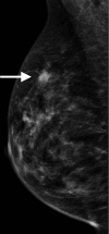
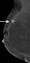
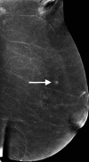
Images in 52-year-old woman with a 1.6-cm, node-negative, grade-1 estrogen receptor–positive/progesterone receptor–positive/human epidermal growth factor receptor 2–negative invasive ductal cancer evident on both the low-energy and iodine images. (a) An asymmetry (arrow) is in the superior right breast on the right mediolateral oblique view. (b) This focal asymmetry is enhanced (arrow) after contrast agent administration on the mediolateral oblique iodine image. (c) Targeted US helped to identify a sonographic correlate of an irregular 1.1-cm mass (arrow) at the 11:00 axis. Subsequent biopsy yielded invasive ductal carcinoma.
Figure 2b:
Figure 2b:
Images in 52-year-old woman with a 1.6-cm, node-negative, grade-1 estrogen receptor–positive/progesterone receptor–positive/human epidermal growth factor receptor 2–negative invasive ductal cancer evident on both the low-energy and iodine images. (a) An asymmetry (arrow) is in the superior right breast on the right mediolateral oblique view. (b) This focal asymmetry is enhanced (arrow) after contrast agent administration on the mediolateral oblique iodine image. (c) Targeted US helped to identify a sonographic correlate of an irregular 1.1-cm mass (arrow) at the 11:00 axis. Subsequent biopsy yielded invasive ductal carcinoma.
Figure 2c:
Figure 2c:
Images in 52-year-old woman with a 1.6-cm, node-negative, grade-1 estrogen receptor–positive/progesterone receptor–positive/human epidermal growth factor receptor 2–negative invasive ductal cancer evident on both the low-energy and iodine images. (a) An asymmetry (arrow) is in the superior right breast on the right mediolateral oblique view. (b) This focal asymmetry is enhanced (arrow) after contrast agent administration on the mediolateral oblique iodine image. (c) Targeted US helped to identify a sonographic correlate of an irregular 1.1-cm mass (arrow) at the 11:00 axis. Subsequent biopsy yielded invasive ductal carcinoma.
Figure 3a:
Figure 3a:
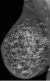
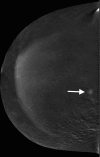
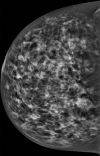
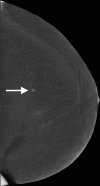
Images in a 65-year-old woman with a 0.8-cm, node-negative, grade 2, estrogen receptor–positive/progesterone receptor–positive/human epidermal growth factor receptor 2–negative invasive ductal cancer only evident at contrast-enhanced digital mammography (CEDM) because of contrast enhancement. No abnormality was depicted on the (a) right mediolateral oblique and (d) craniocaudal views from low-energy images of the screening CEDM. A 5-mm enhanced mass (arrows in b and c) is visible only after contrast administration on the (b) mediolateral oblique and (c) craniocaudal iodine images. Subsequent biopsy yielded invasive ductal carcinoma.
Figure 3b:
Figure 3b:
Images in a 65-year-old woman with a 0.8-cm, node-negative, grade 2, estrogen receptor–positive/progesterone receptor–positive/human epidermal growth factor receptor 2–negative invasive ductal cancer only evident at contrast-enhanced digital mammography (CEDM) because of contrast enhancement. No abnormality was depicted on the (a) right mediolateral oblique and (d) craniocaudal views from low-energy images of the screening CEDM. A 5-mm enhanced mass (arrows in b and c) is visible only after contrast administration on the (b) mediolateral oblique and (c) craniocaudal iodine images. Subsequent biopsy yielded invasive ductal carcinoma.
Figure 3c:
Figure 3c:
Images in a 65-year-old woman with a 0.8-cm, node-negative, grade 2, estrogen receptor–positive/progesterone receptor–positive/human epidermal growth factor receptor 2–negative invasive ductal cancer only evident at contrast-enhanced digital mammography (CEDM) because of contrast enhancement. No abnormality was depicted on the (a) right mediolateral oblique and (d) craniocaudal views from low-energy images of the screening CEDM. A 5-mm enhanced mass (arrows in b and c) is visible only after contrast administration on the (b) mediolateral oblique and (c) craniocaudal iodine images. Subsequent biopsy yielded invasive ductal carcinoma.
Figure 3d:
Figure 3d:
Images in a 65-year-old woman with a 0.8-cm, node-negative, grade 2, estrogen receptor–positive/progesterone receptor–positive/human epidermal growth factor receptor 2–negative invasive ductal cancer only evident at contrast-enhanced digital mammography (CEDM) because of contrast enhancement. No abnormality was depicted on the (a) right mediolateral oblique and (d) craniocaudal views from low-energy images of the screening CEDM. A 5-mm enhanced mass (arrows in b and c) is visible only after contrast administration on the (b) mediolateral oblique and (c) craniocaudal iodine images. Subsequent biopsy yielded invasive ductal carcinoma.
Figure 4a:
Figure 4a:
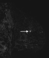
Images in a 64-year-old woman with results that were false-positive for cancer at contrast-enhanced digital mammography. An enhancing 0.5-cm mass (arrows) is seen in the left upper outer quadrant on the (a) mediolateral oblique and (b) craniocaudal iodine views. No abnormality was observed on the low-energy images or at targeted US. (c) Subtraction image from MRI demonstrates a correlate (arrow). MRI-guided biopsy yielded fibroadenomatoid changes and other benign pathologic results.
Figure 4b:
Figure 4b:
Images in a 64-year-old woman with results that were false-positive for cancer at contrast-enhanced digital mammography. An enhancing 0.5-cm mass (arrows) is seen in the left upper outer quadrant on the (a) mediolateral oblique and (b) craniocaudal iodine views. No abnormality was observed on the low-energy images or at targeted US. (c) Subtraction image from MRI demonstrates a correlate (arrow). MRI-guided biopsy yielded fibroadenomatoid changes and other benign pathologic results.
Figure 4c:
Figure 4c:
Images in a 64-year-old woman with results that were false-positive for cancer at contrast-enhanced digital mammography. An enhancing 0.5-cm mass (arrows) is seen in the left upper outer quadrant on the (a) mediolateral oblique and (b) craniocaudal iodine views. No abnormality was observed on the low-energy images or at targeted US. (c) Subtraction image from MRI demonstrates a correlate (arrow). MRI-guided biopsy yielded fibroadenomatoid changes and other benign pathologic results.

Comment in

References

    1. Smith RA, Duffy SW, Gabe R, Tabar L, Yen AM, Chen TH. The randomized trials of breast cancer screening: what have we learned? Radiol Clin North Am 2004;42(5):793–806, v. - PubMed
    1. Tabár L, Vitak B, Chen TH, et al. . Swedish two-county trial: impact of mammographic screening on breast cancer mortality during 3 decades. Radiology 2011;260(3):658–663. - PubMed
    1. Humphrey LL, Helfand M, Chan BK, Woolf SH. Breast cancer screening: a summary of the evidence for the U.S. Preventive Services Task Force. Ann Intern Med 2002;137(5 Part 1):347–360. - PubMed
    1. Tilanus-Linthorst M, Verhoog L, Obdeijn IM, et al. . A BRCA1/2 mutation, high breast density and prominent pushing margins of a tumor independently contribute to a frequent false-negative mammography. Int J Cancer 2002;102(1):91–95. [Published correction appears in Int J Cancer 2002;102(6):665.] 10.1002/ijc.10666 - DOI - PubMed
    1. Berg WA, Blume JD, Cormack JB, et al. . Combined screening with ultrasound and mammography vs mammography alone in women at elevated risk of breast cancer. JAMA 2008;299(18):2151–2163 [Published correction appears in JAMA 2010;303(15):1482.] 10.1001/jama.299.18.2151 - DOI - PMC - PubMed

Publication types