. 2019 Nov;23(11):7525-7534.
doi: 10.1111/jcmm.14621. Epub 2019 Aug 27.

NOV/CCN3 induces cartilage protection by inhibiting PI3K/AKT/mTOR pathway

Affiliations

NOV/CCN3 induces cartilage protection by inhibiting PI3K/AKT/mTOR pathway

Xiaojian Huang et al. J Cell Mol Med. 2019 Nov.

Abstract

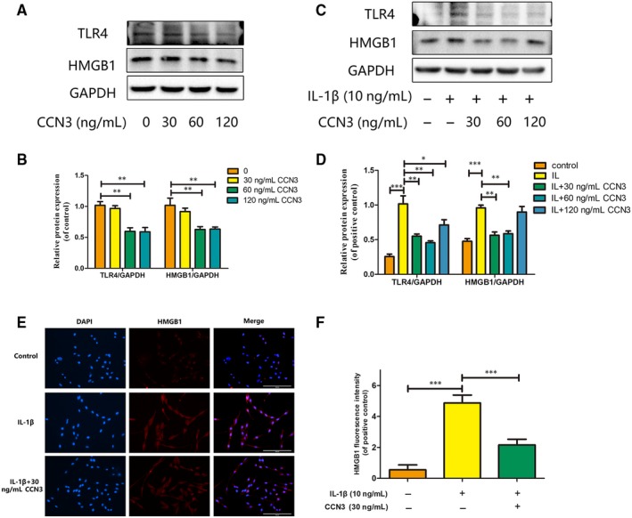
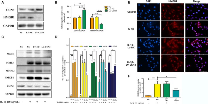
Osteoarthritis (OA), an age-related degenerative joint disease, is pathologically characterized by articular cartilage degeneration and synovial inflammation. Nephroblastoma overexpressed (NOV or CCN3), a matricellular protein, is a primary member of the CCN family (Cyr61, Ctgf, NOV) of proteins and is involved in various inflammatory disorders. Previous studies reported that CCN3 might play a therapeutic role in OA. However, the underlying mechanism remains unclear. In this study, we confirmed the expression of CCN3 was decreased in human and rat OA articular cartilage. Recombinant CCN3 ameliorated the IL-1β-induced matrix catabolism, as demonstrated by MMP1, MMP3, MMP13, ADAMTS5 and iNOS expression, in vitro. In addition, the degradation of cartilage matrix such as collagen 2 and aggrecan could be reversed by CCN3. Furthermore, we found CCN3 promoted autophagy as Atg5, Beclin1 and LC3-II expression were increased. High-mobility group box 1 was negatively correlated with CCN3 in IL-1β-induced osteoarthritis responses, and HMGB1 is involved in the protective effect of CCN3 in OA. Moreover, CCN3 overexpression decreased the expression of HMGB1 and reversed the IL-1β induced MMPs production. Additionally, recombinant CCN3 or CCN3 overexpression attenuated the activation of PI3K/AKT/mTOR pathway induced by IL-1β. Our study presents new mechanisms of CCN3 in osteoarthritis and indicates that CCN3 can serve as a novel potential therapeutic target for osteoarthritis.

Keywords: CCN3; HMGB1; IL-1β; MMPs; osteoarthritis.


Conflict of interest statement

The authors confirm that there is no conflict of interest.

Figures

Figure 1
Figure 1
CCN3 expression is decreased in human knee OA articular cartilage. A, Safranin‐O and fast green staining showed significant cartilage erosion in human OA cartilage compared with normal cartilage. Immunohistochemistry showed decreased CCN3 expression in human OA cartilage. B, Quantitative analysis showed lower CCN3 expression in OA cartilage than in healthy cartilage (n = 6). C, Western blot and quantitative analysis showed lower CCN3 expression in SW1353 cells stimulated with IL‐1β for 24 h than in normal cells. (original magnification × 40, scale bar: 100 μm). Significant differences between groups are indicated as ***P < .001, **P < .01 and *P < .05
Figure 2
Figure 2
CCN3 expression is decreased in rat knee OA articular cartilage. A, Safranin‐O and fast green staining showed significant cartilage erosion in rat OA cartilage compared with normal cartilage. Immunohistochemistry showed decreased CCN3 expression in rat OA cartilage. B, Quantitative analysis showed lower CCN3 expression in OA cartilage than in normal cartilage (n = 6). C, Western blot and quantitative analysis showed lower CCN3 expression in rat chondrocytes stimulated with IL‐1β for 24 h than in normal cells. (original magnification × 40, scale bar: 100 μm). Data represent mean ± SD of three independent experiments, each done in triplicate. Significant differences between groups are indicated as ***P < .001, **P < .01 and *P < .05
Figure 3
Figure 3
CCN3 ameliorates the IL‐1β‐Induced catabolism, cartilage matrix degradation and promotes autophagy process in vitro. A, SW1353 cells were treated by IL‐1β at different doses for 24 h. Western blotting was performed to examine changes in MMPs, ADAMTS5, iNOS, collagen 2 and aggrecan expression. B, Quantification of MMP, ADAMTS5, iNOS, collagen 2 and aggrecan immunoblots. C, SW1353 cells were treated with recombinant CCN3 (30, 60 and 120 ng/mL) and IL‐1β (10 ng/mL) for 24 h. Western blotting was performed to examine changes in MMPs, ADAMTS5, iNOS, collagen 2 and aggrecan expression. D, Quantification of MMP, ADAMTS5, iNOS, collagen 2 and aggrecan immunoblots. E, SW1353 cells were treated with recombinant CCN3 (30, 60 ng/mL) and IL‐1β (10 ng/mL) for 24 h. Western blotting was performed to examine changes in Atg5, Beclin1 and LC3‐I/II expression. F, Quantification of Atg5, Beclin1 and LC3‐I/II immunoblots. Data represent mean ± SD of three independent experiments, each done in triplicate. Significant differences between groups are indicated as ***P < .001, **P < .01 and *P < .05
Figure 4
Figure 4
HMGB1 is negatively correlated with CCN3 in IL‐1β induced osteoarthritis responses. A, HE staining showed significant cartilage erosion in human OA cartilage compared with normal cartilage. Immunohistochemistry showed higher HMGB1 expression in human OA cartilage than in normal cartilage. (original magnification × 40, scale bar: 100 μm) B, SW1353 cells were treated with IL‐1β (10 ng/mL) at different time points. Western blot was performed to examine changes in TLR4, HMGB1 and CCN3 expression. C, Quantitative analysis showed HMGB1 is negatively correlated with CCN3 in IL‐1β treated SW1353 cells in a time‐dependent manner. D, SW1353 cells were treated with IL‐1β at different dose points for 24 h. Western blot was performed to examine changes in HMGB1 and CCN3 expression. E, Quantitative analysis showed HMGB1 is negatively correlated with CCN3 in IL‐1β‐induced osteoarthritis responses in a dose‐dependent manner. The data represent mean ± SD of three independent experiments, each done in triplicate. Significant differences between groups are indicated as ***P < .001, **P < .01 and *P < .05
Figure 5
Figure 5
HMGB1 is involved in the protective effect of CCN3 in osteoarthritis. A, SW1353 cells were treated by recombinant CCN3 (30, 60 and 120 ng/mL) for 24 h. Western blot was performed to examine changes in TLR4 and HMGB1 expression. B, Quantitative analysis showed that recombinant CCN3 dose‐dependent decreased HMGB1 and TLR4 expression. C, SW1353 cells were treated with recombinant CCN3 (30, 60 and 120 ng/mL) and IL‐1β (10 ng/mL) for 24 h. Western blot was performed to examine changes in HMGB1 and CCN3 expression. D, Quantitative analysis showed recombinant CCN3 dose‐dependent decreased the expression of HMGB1 as well as TLR4. D, Quantitative analysis showed CCN3 (30, 60 ng/mL) reversed the increased HMGB1 and TLR4 production induced by IL‐1β. E, SW1353 cells were treated with 30 ng/mL recombinant CCN3 and IL‐1β (10 ng/mL) for 24 h. Immunofluorescence showed CCN3 reduced the amount of HMGB1. F, The fluorescence intensity of HMGB1 was quantified. Data represent mean ± SD of three independent experiments, each done in triplicate. Significant differences between groups are indicated as ***P < .001, **P < .01 and *P < .05
Figure 6
Figure 6
CCN3 overexpression decreases the expression of HMGB1 and reverses the IL‐1β induced MMPs. A, CCN3 was overexpressed via transfection of lenti‐CCN3. Western blot was performed to examine changes in CCN3 and HMGB1 expression. B, Quantitative analysis showed CCN3 overexpression could decrease the HMGB1 expression. C, CCN3 overexpression cells were treated with IL‐1β (10 ng/mL) for 24 h. Western blot showed CCN3 overexpression attenuated the increased MMP1, MMP3, MMP13 and HMGB1 expression induced by IL‐1β. D, Quantification of MMP1, MMP3, MMP13, HMGB1 and CCN3 immunoblots E, Immunofluorescence showed CCN3 overexpression reduced the amount of HMGB1. F, The fluorescence intensity of HMGB1 was quantified. Data represent mean ± SD of three independent experiments, each performed in triplicate. Significant differences between groups are indicated as ***P < .001, **P < .01 and *P < .05
Figure 7
Figure 7
CCN3 induces cartilage protection via blocking the PI3K/AKT/mTOR pathway. A, SW1353 cells were treated with recombinant CCN3 (30, 60 ng/mL) and IL‐1β (10 ng/mL) for 30 min. Western blot showed recombinant CCN3 attenuated the increased protein levels of P‐PI3K, P‐AKT and P‐mTOR activated by IL‐1β. B, Quantification of P‐PI3K, P‐AKT and P‐mTOR immunoblots. C, CCN3 overexpression cells were treated IL‐1β (10 ng/mL) for 30 min. Western blot showed CCN3 overexpression attenuated the increased protein levels of P‐PI3K, P‐AKT and P‐mTOR activated by IL‐1β. D, Quantification of P‐PI3K, P‐AKT and P‐mTOR immunoblots. The data represent the mean ± SD of three independent experiments, each performed in triplicate. Significant differences between groups are indicated as ***P < .001, **P < .01 and *P < .05

References

    1. Tong W, Zeng Y, Chow D, et al. Wnt16 attenuates osteoarthritis progression through a PCP/JNK‐mTORC1‐PTHrP cascade. Ann Rheum Dis. 2019;78:551‐561. - PubMed
    1. Xing D, Xu Y, Liu Q, et al. Osteoarthritis and all‐cause mortality in worldwide populations: grading the evidence from a meta‐analysis. Sci Rep. 2016;6:24393. - PMC - PubMed
    1. O'Neill TW, McCabe PS, McBeth J. Update on the epidemiology, risk factors and disease outcomes of osteoarthritis. Best Pract Res Clin Rheumatol. 2018;32:312‐326. - PubMed
    1. Mandl LA. Osteoarthritis year in review 2018: clinical. Osteoarthritis Cartilage. 2019;27:359‐364. - PubMed
    1. Verma P, Dalal K. ADAMTS‐4 and ADAMTS‐5: key enzymes in osteoarthritis. J Cell Biochem. 2011;112:3507‐3514. - PubMed

Publication types