. 2019 Aug 26;20(17):4164.
doi: 10.3390/ijms20174164.

β-Glucose-1,6-Bisphosphate Stabilizes Pathological Phophomannomutase2 Mutants In Vitro and Represents a Lead Compound to Develop Pharmacological Chaperones for the Most Common Disorder of Glycosylation, PMM2-CDG

Affiliations

β-Glucose-1,6-Bisphosphate Stabilizes Pathological Phophomannomutase2 Mutants In Vitro and Represents a Lead Compound to Develop Pharmacological Chaperones for the Most Common Disorder of Glycosylation, PMM2-CDG

Maria Monticelli et al. Int J Mol Sci. .

Abstract

A large number of mutations causing PMM2-CDG, which is the most frequent disorder of glycosylation, destabilize phosphomannomutase2. We looked for a pharmacological chaperone to cure PMM2-CDG, starting from the structure of a natural ligand of phosphomannomutase2, α-glucose-1,6-bisphosphate. The compound, β-glucose-1,6-bisphosphate, was synthesized and characterized via 31P-NMR. β-glucose-1,6-bisphosphate binds its target enzyme in silico. The binding induces a large conformational change that was predicted by the program PELE and validated in vitro by limited proteolysis. The ability of the compound to stabilize wild type phosphomannomutase2, as well as frequently encountered pathogenic mutants, was measured using thermal shift assay. β-glucose-1,6-bisphosphate is relatively resistant to the enzyme that specifically hydrolyses natural esose-bisphosphates.

Keywords: PMM2-CDG; glucose-1,6-bisphosphate; pharmacological chaperone.


Conflict of interest statement

The authors declare no conflict of interest.

Figures

Figure 1
Figure 1
31P-NMR characterization of α-D-Glucose 1,6-bisphosphate (αG16P) and β-glucose-1,6-bisphosphate (βG16P). (A) 1H-decoupled one-dimensional 31P spectrum. (B,C) Selected regions of the 1H-31P HSQC spectrum. Both the spectra were acquired in H2O + D2O 10% in the presence of EDTA 50 mM; ppm were referred to creatine phosphate (0 ppm, 31P scale) and to trimethylsilylpropanoic acid (0 ppm, 1H scale). P(6) and P(1) indicate the positions of phosphorous nuclei in the molecules; §: inorganic phosphate.
Figure 2
Figure 2
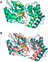
In silico binding models for PMM2 and βG16P: either the phosphate proximal to C6, P6_Mg mode (A), or the phosphate proximal to C1, P1_Mg mode (B), can interact with the catalytic site. PMM2 in the initial state is in cyan (A,B); the two closed conformations are in green (A) and pink (B); βG16P is red; Mg2+ are shown as spheres.
Figure 3
Figure 3
Interactions between PMM2 and ligands: residues that interact with βG16P by hydrogen bonds or salt bridges in P1_Mg mode (A) and P6_Mg mode (B) respectively; residues that interact with αG16P by hydrogen bonds or salt bridges in P1_Mg (C) mode and P6_Mg mode (D) respectively.
Figure 4
Figure 4
Limited proteolysis of wild type PMM2 by trypsin. Wild type PMM2 was incubated at 37 °C with trypsin in a 50:1 ratio in the presence or the absence of βG16P or αG16P 0.5 mM. Aliquots were withdrawn at specified times (0, 20, 60, 120 min) and analyzed by SDS-PAGE and Coomassie staining.
Figure 5
Figure 5
PMM2 phosphoglucomutase activity monitored by fluorescence spectroscopy (A) and phosphomannomutase activity monitored by 31P-NMR (B). (A) A total of 0.08 μg PMM2 were incubated at room temperature with different combinations of α or βG1P as substrates and α or βG16P as activators, in the presence of G6PDH and NADP+. (B) A total of 0.12 μg PMM2 were incubated at 32 °C with 1 mM α-mannose-1-phosphate (αM1P); αG16P 5 μM was used as an activator, in the absence or the presence of βG16P 5 or 50 μM.
Figure 6
Figure 6
Thermal stability in the presence of ligands. wt-PMM2 0.3 mg/mL was incubated with different ligands 0.5 mM ((A) αG16P and βG16P; (B) αG1P and βG1P, with and without vanadate); the melting curves were measured in the presence of dithiothreitol (DTT) 1 mM and Sypro Orange 2.4x, from 20 to 90 °C with increments of 1 °C/min.
Figure 7
Figure 7
Thermal stability of pathological mutants in the presence of ligands. F119L-PMM2 (A) and V129M-PMM2 (B) 0.3 mg/mL were incubated with different ligands, αG16P (0.5 mM), and βG16P (0.5 mM). The melting curves were measured in the presence of DTT 1 mM and Sypro Orange 2.4x, from 20 to 90 °C with increments of 1 °C/min.

References

    1. Pirard M., Achouri Y., Collet J.F., Schollen E., Matthijs G., Van Schaftingen E. Kinetic properties and tissular distribution of mammalian phosphomannomutase isozymes. Biochem. J. 1999;339:201–207. doi: 10.1042/bj3390201. - DOI - PMC - PubMed
    1. Veiga-da-Cunha M., Vleugels W., Maliekal P., Matthijs G., Van Schaftingen E. Mammalian phosphomannomutase PMM1 is the brain IMP-sensitive glucose-1,6-bisphosphatase. J. Biol. Chem. 2008;283:33988–33993. doi: 10.1074/jbc.M805224200. - DOI - PMC - PubMed
    1. Citro V., Cimmaruta C., Liguori L., Viscido G., Cubellis M.V., Andreotti G. A mutant of phosphomannomutase1 retains full enzymatic activity, but is not activated by IMP: Possible implications for the disease PMM2-CDG. PLoS ONE. 2017;12:e0189629. doi: 10.1371/journal.pone.0189629. - DOI - PMC - PubMed
    1. Van Schaftingen E., Jaeken J. Phosphomannomutase deficiency is a cause of carbohydrate-deficient glycoprotein syndrome type I. FEBS Lett. 1995;377:318–320. - PubMed
    1. Kjaergaard S., Skovby F., Schwartz M. Carbohydrate-deficient glycoprotein syndrome type 1A: Expression and characterisation of wild type and mutant PMM2 in E. coli. Eur. J. Hum. Genet. 1999;7:884–888. doi: 10.1038/sj.ejhg.5200398. - DOI - PubMed

MeSH terms

Substances

Supplementary concepts

LinkOut - more resources