Review
. 2019 Aug 26;8(9):976.
doi: 10.3390/cells8090976.

Physiology, Pathology and Regeneration of Salivary Glands

Affiliations
Review

Physiology, Pathology and Regeneration of Salivary Glands

Cristina Porcheri et al. Cells. .

Abstract

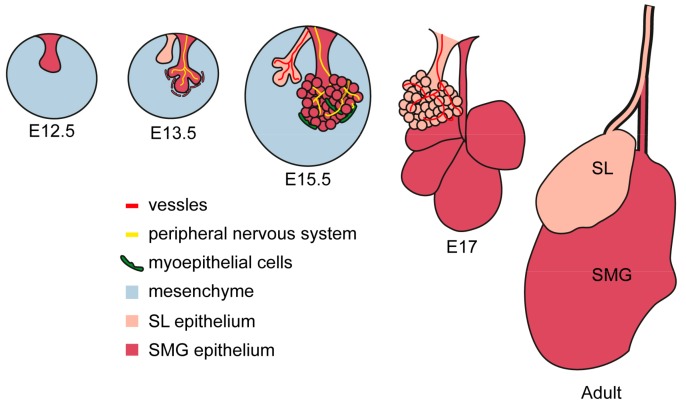
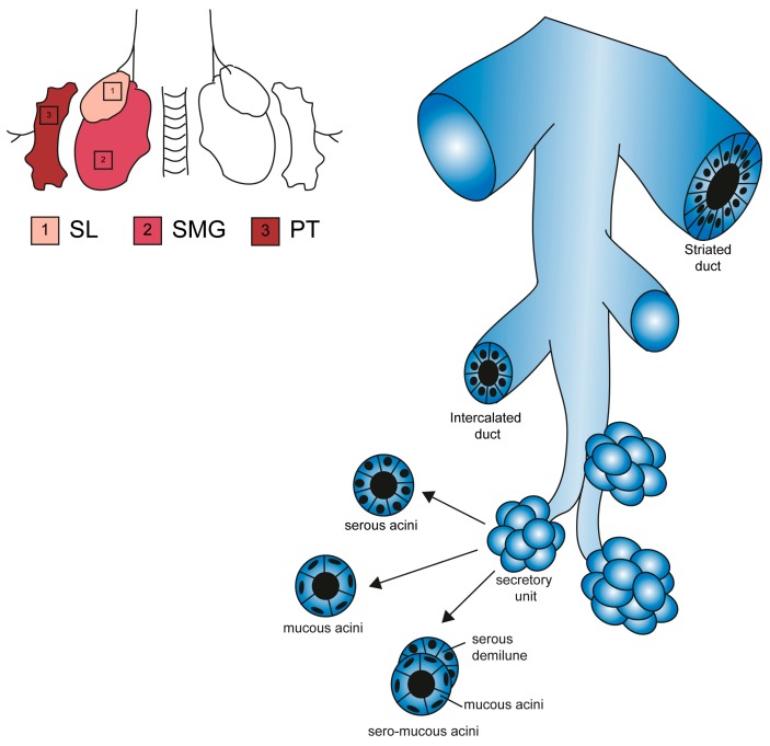
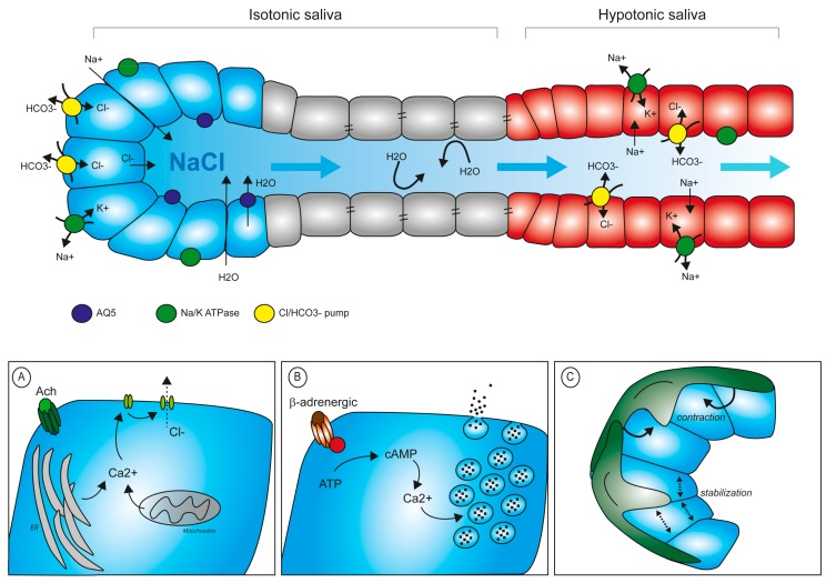
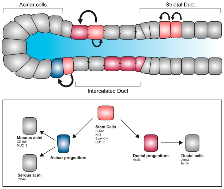
Salivary glands are essential structures in the oral cavity. A variety of diseases, such as cancer, autoimmune diseases, infections and physical traumas, can alter the functionality of these glands, greatly impacting the quality of life of patients. To date, no definitive therapeutic approach can compensate the impairment of salivary glands, and treatment are purely symptomatic. Understanding the cellular and molecular control of salivary glands function is, therefore, highly relevant for therapeutic purposes. In this review, we provide a starting platform for future studies in basic biology and clinical research, reporting classical ideas on salivary gland physiology and recently developed technology to guide regeneration, reconstruction and substitution of the functional organs.

Keywords: exocrine glands; oral epithelium; salivary gland-resident stem cells; salivary glands; xerostomia.


Conflict of interest statement

The authors declare no conflicts of interest.

Figures

Figure 1
Figure 1
Embryonic development of murine SMG and SL glands. Embryonic development starts with an epithelial placode, which, in turn, invades the mesenchymal layer and branches up. Innervation and vascularization advance in parallel with the growth of salivary glands primordia and directly regulates its maturation. Myoepithelial cells arise from the external epithelial layer and develop into the contracting unit involved in the regulation of secretion.
Figure 2
Figure 2
Structural features of major salivary glands. Schematic representation of SL, SMG and PG glands and their functional elements [35]. All glands contain ducts of bigger diameter (striated duct) and branching ducts (intercalated ducts). The acinar end-pieces are secretory units specialized in a single type of secretion (serous, mucous or mixed).
Figure 3
Figure 3
Saliva production and regulation of secretion. Composition of primary saliva and its progressive conversion into a hypotonic fluid. Primary saliva is rich in electrolytes which attract water into the lumen. In a secondary step, electrolytes are resorbed via the epithelial duct cells to reduce their concentration in the saliva before release in the oral cavity. (A) Schematic representation of intracellular Ca2+ control levels upon muscarinic stimuli. (B) Secretion of proteins into the lumen requires Ca2+-mediated degranulation upon β-adrenergic stimulation. (C) Myoepithelial cells participate in the mechanical contraction stimulating secretion and in the stabilization of acinar structure during the resting phase.
Figure 4
Figure 4
Stem cell pool and regeneration of salivary glands. Salivary glands contain several pools of undifferentiated progenitors able to self-renew, proliferate and give rise to differentiated acinar and ductal cells. Several markers allow the identification of stem cells and committed progenitors, allowing fate tracking and molecular discrimination.

Similar articles

Cited by

References

    1. Amano O., Mizobe K., Bando Y., Sakiyama K. Anatomy and histology of rodent and human major salivary glands: Overview of the Japan salivary gland society-sponsored workshop. Acta Histochem. Cytochem. 2012;45:241–250. doi: 10.1267/ahc.12013. - DOI - PMC - PubMed
    1. Dobrosielski-Vergona K. Biology of the Salivary Glands. CRC Press, Taylor & Francis Group; Abingdon, UK: 1993.
    1. Korsrud F.R., Brandtzaeg P. Quantitative immunohistochemistry of immunoglobulin- and J-chain-producing cells in human parotid and submandibular salivary glands. Immunology. 1980;39:129–140. - PMC - PubMed
    1. Smith D.J., Taubman M.A., King W.F. Immunological features of minor salivary gland saliva. J. Clin. Immunol. 1987;7:449–455. doi: 10.1007/BF00915054. - DOI - PubMed
    1. Treuting P.M., Dintzis S.M., Frevert C.W., Liggitt D., Liggitt H.D., Montine K.S. Comparative Anatomy and Histology: A Mouse and Human Atlas (Expert Consult) Elsevier Inc.; UK: 2012. [(accessed on 3 August 2019)]. Available online: https://books.google.ch/books?id=Bqn23_270Q8C.

Publication types

LinkOut - more resources