Case Reports
. 2019 Aug 27;7(1):138.
doi: 10.1186/s40478-019-0781-8.

Clinical, histological, and genetic characterization of PYROXD1-related myopathy

Affiliations
Case Reports

Clinical, histological, and genetic characterization of PYROXD1-related myopathy

Xavière Lornage et al. Acta Neuropathol Commun. .

Abstract

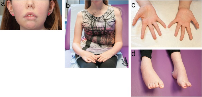
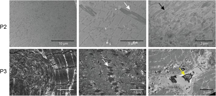
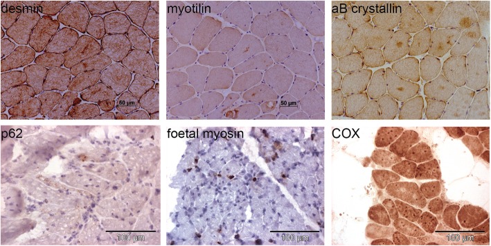
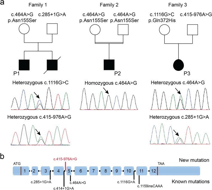
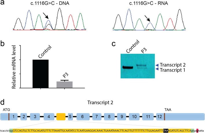
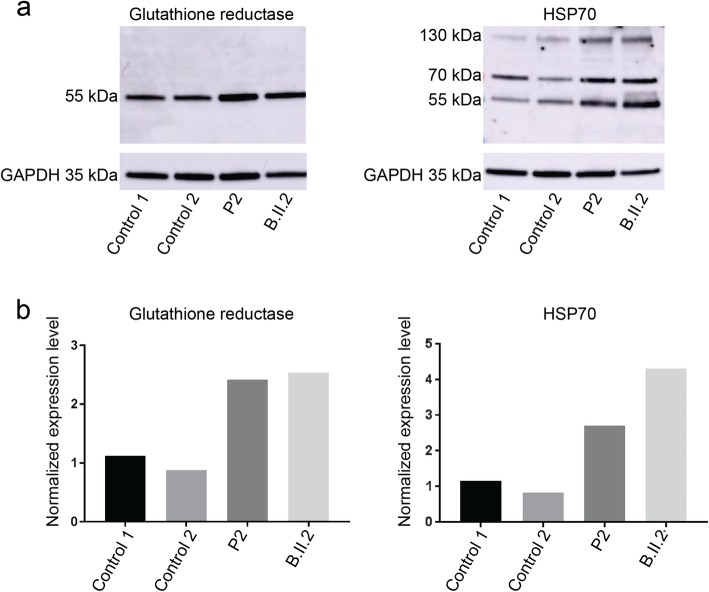
Recessive mutations in PYROXD1, encoding an oxidoreductase, were recently reported in families with congenital myopathy or limb-girdle muscular dystrophy. Here we describe three novel PYROXD1 families at the clinical, histological, and genetic level. Histological analyses on muscle biopsies from all families revealed fiber size variability, endomysial fibrosis, and muscle fibers with multiple internal nuclei and cores. Further characterization of the structural muscle defects uncovered aggregations of myofibrillar proteins, and provided evidence for enhanced oxidative stress. Sequencing identified homozygous or compound heterozygous PYROXD1 mutations including the first deep intronic mutation reinforcing a cryptic donor splice site and resulting in mRNA instability through exonisation of an intronic segment. Overall, this work expands the PYROXD1 mutation spectrum, defines and specifies the histopathological hallmarks of the disorder, and indicates that oxidative stress contributes to the pathomechanism. Comparison of all new and published cases uncovered a genotype/phenotype correlation with a more severe and early-onset phenotypic presentation of patients harboring splice mutations resulting in reduced PYROXD1 protein levels compared with patients carrying missense mutations.

Keywords: Congenital myopathy; LGMD; Myofibrillar inclusions; Oxidoreductase; PYROXD1.


Conflict of interest statement

The authors declare that they have no competing interests.

Figures

Fig. 1
Fig. 1
Photographs of P3. a Low-set ears, b scoliosis, c hand size asymmetry, d arched feet
Fig. 2
Fig. 2
Skeletal muscle histopathology. H&E, NADH-TR, and Gomori trichrome staining of transverse muscle section from P1, P2, and P3 revealed similar histological features as fiber-size heterogeneity, fibrosis, rods, and fibers with multiple internalized nuclei (black arrows) and cores (white arrows)
Fig. 3
Fig. 3
Skeletal muscle ultrastructure. Electron microscopy on muscle section from P2 and P3 confirmed the presence of cores and rods (white arrows), and revealed glycogen accumulations (black arrow), abnormal mitochondria, and dense osmiophilic bodies (yellow arrow) of unknown origin outside the sarcolemma and within fibres
Fig. 4
Fig. 4
Protein accumulations in patient muscles. Immuno- and chemical staining of muscle biopsies from P2 and P3 revealed accumulations of the myofibrillar proteins desmin, myotilin, and alpha B crystallin, and of the mitochondrial marker COX, and detected a few fibers expressing foetal myosin, or with positive labelling for the p62 autophagy marker
Fig. 5
Fig. 5
Identification of PYROXD1 mutations. a Pedigrees of three novel PYROXD1 families and chromatopherograms showing the mutations. b Schematic representation of PYROXD1 and position of known mutations (black) and the novel mutation (red)
Fig. 6
Fig. 6
Characterization of the deep intronic mutation. a The c.1116G > C mutation appears heterozygous on the P3 DNA and homozygous on the RNA. b The PYROXD1 mRNA was strongly reduced in the muscle from P3. c Discriminative PCR on skeletal muscle cDNA revealed the presence of an aberrant amplicon with increased size (transcript 2). d Sequencing of the aberrant transcript 2 showed the inclusion of an additional 110 nt exon with in-frame stop codon (highlighted in black). The intronic mutation (red) reinforces a cryptic donor site (green)
Fig. 7
Fig. 7
Increased oxidative stress markers. a Western blot and b quantification on muscle extracts from two PYROXD1 patients revealed increased protein levels of HSP70 monomers (70 kDa) and dimers (140 kDa) and glutathione reductase compared with age-matched controls

References

    1. Dubowitz Victor, Sewry Caroline A. Muscle Biopsy. 2007. Normal muscle; pp. 41–74.
    1. Gonorazky HD, Bonnemann CG, Dowling JJ. The genetics of congenital myopathies. Handb Clin Neurol. 2018;148:549–564. doi: 10.1016/B978-0-444-64076-5.00036-3. - DOI - PubMed
    1. Ji LL, Fu R. Responses of glutathione system and antioxidant enzymes to exhaustive exercise and hydroperoxide. J Appl Physiol (1985) 1992;72:549–554. doi: 10.1152/jappl.1992.72.2.549. - DOI - PubMed
    1. Maquat LE. Nonsense-mediated mRNA decay: splicing, translation and mRNP dynamics. Nat Rev Mol Cell Biol. 2004;5:89–99. doi: 10.1038/nrm1310. - DOI - PubMed
    1. Nance JR, Dowling JJ, Gibbs EM, Bonnemann CG. Congenital myopathies: an update. Curr Neurol Neurosci Rep. 2012;12:165–174. doi: 10.1007/s11910-012-0255-x. - DOI - PMC - PubMed

Publication types

Substances