. 2019 Oct 18;294(42):15386-15394.
doi: 10.1074/jbc.RA119.009000. Epub 2019 Aug 27.

The mammalian host protein DAP5 facilitates the initial round of translation of Coxsackievirus B3 RNA

Affiliations

The mammalian host protein DAP5 facilitates the initial round of translation of Coxsackievirus B3 RNA

Pratik Dave et al. J Biol Chem. .

Abstract

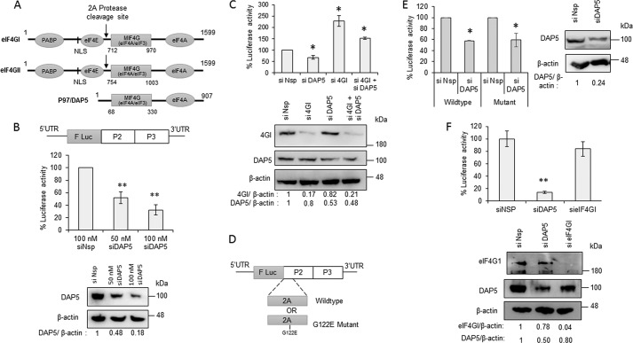
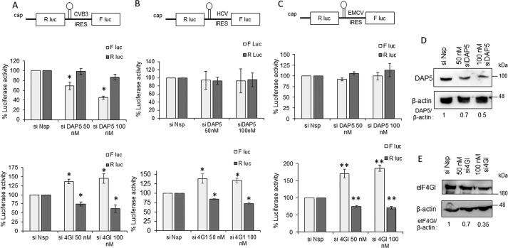
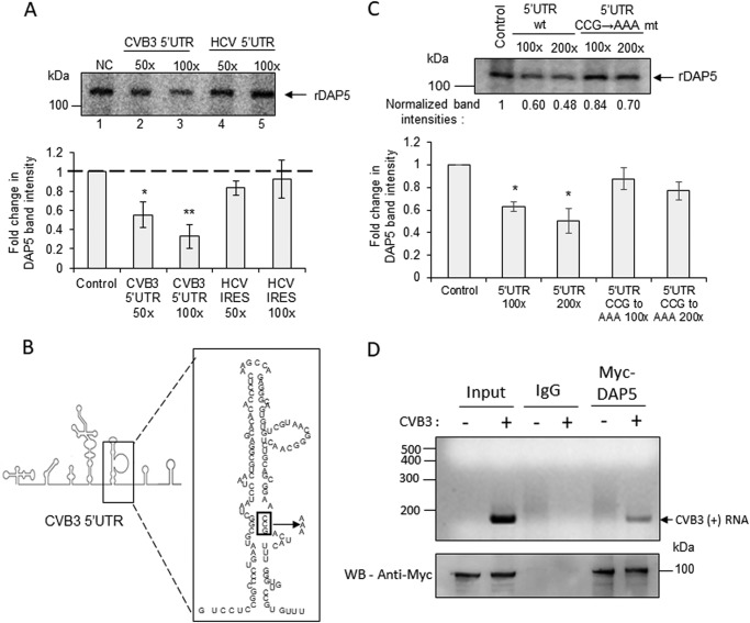
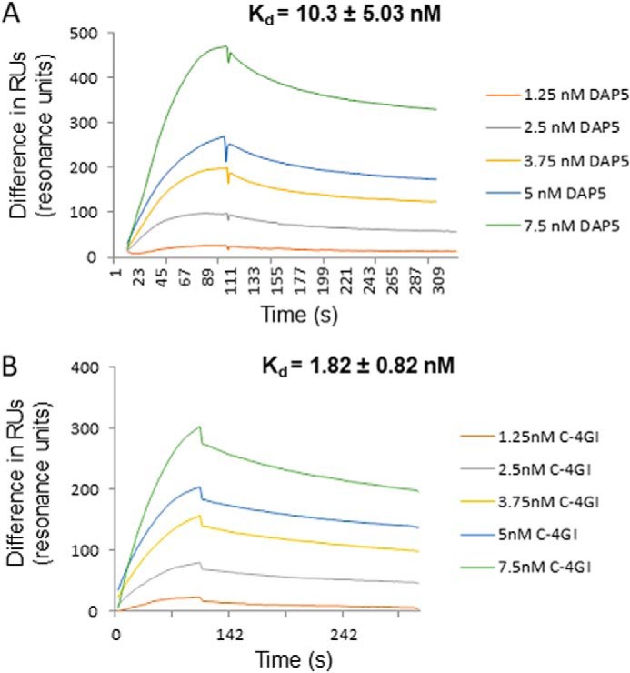
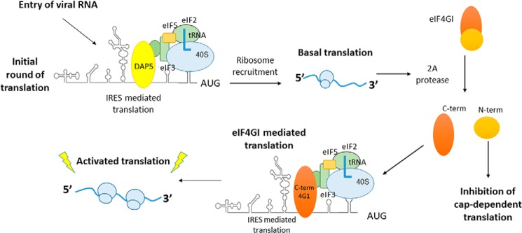
During enteroviral infections, the canonical translation factor eukaryotic translation initiation factor 4 γ I (eIF4GI) is cleaved by viral protease 2A. The resulting C-terminal fragment is recruited by the viral internal ribosome entry site (IRES) for efficient translation of the viral RNA. However, the 2A protease is not present in the viral capsid and is synthesized only after the initial round of translation. This presents the conundrum of how the initial round of translation occurs in the absence of the C-terminal eIF4GI fragment. Interestingly, the host protein DAP5 (also known as p97, eIF4GIII, and eIF4G2), an isoform of eIF4GI, closely resembles the eIF4GI C-terminal fragment produced after 2A protease-mediated cleavage. Using the Coxsackievirus B3 (CVB3) IRES as a model system, here we demonstrate that DAP5, but not the full-length eIF4GI, is required for CVB3 IRES activity for translation of input viral RNA. Additionally, we show that DAP5 is specifically required by type I IRES but not by type II or type III IRES, in which cleavage of eIF4GI has not been observed. We observed that both DAP5 and C-terminal eIF4GI interact with CVB3 IRES in the same region, but DAP5 exhibits a lower affinity for CVB3 IRES compared with the C-terminal eIF4GI fragment. It appears that DAP5 is required for the initial round of viral RNA translation by sustaining a basal level of CVB3 IRES activity. This activity leads to expression of 2A protease and consequent robust CVB3 IRES-mediated translation by the C-terminal eIF4GI fragment.

Keywords: Coxsackievirus B3; DAP5; IRES mediated translation; RNA virus; eukaryotic translation initiation; eukaryotic translation initiation factor 4G (eIF4G); internal initiation of translation; translation; viral protease.


Conflict of interest statement

The authors declare that they have no conflicts of interest with the contents of this article

Figures

Figure 1.
Figure 1.
DAP5 is required for CVB3 IRES mediated translation. A, schematic of isoforms of eIF4G in mammalian cells. Different domains in individual proteins, the length of the proteins, and the 2A protease cleavage site are indicated. Poly-A binding protein (PABP) interacting domain and Nuclear Localisation Signal (NLS) are indicated in the N-Terminus of the schematic. B, effect of partial silencing of DAP5 on the CVB3 life cycle. Top panel, schematic of CVB3 replicon RNA. F Luc indicated the Firefly Luciferase gene in the CVB3 replicon. CVB3 replicon RNA was transfected in cells transfected previously with the indicated siRNAs, and cells were processed for luciferase activity 8 h after replicon RNA transfection. Center panel, luciferase activity. Bottom panel, Western blot indicating the DAP5 and β-actin protein levels. The band intensities were quantified, and silencing efficiency is represented normalized to β-actin. C, effect of knockdown of DAP5 or eIF4GI or both on the early stage of the CVB3 life cycle. Top panel, luciferase activity indicating translation of the CVB3 IRES. Bottom panel, Western blot showing the DAP5 and β-Actin protein levels upon treatment of siDAP5, sieIF4GI, or both. D, schematic of WT and 2A protease mutant CVB3 replicon RNAs. The G122E mutation in the 2A protease region of the CVB3 replicon RNA is indicated. E, effect of partial silencing of DAP5 on WT and mutant CVB3 replicon RNA translation in early stages. Left panel, luciferase activity indicating translation of the CVB3 IRES. Right panel, Western blotting showing the DAP5 and β-actin protein levels upon siDAP5 treatment. F, effect of partial silencing of DAP5 and eIF4GI on CVB3 IRES activity 1.5 h after CVB3 infection. The CVB3-RLuc virus was used for this experiment, and the top panel represents percentage luciferase activity. All graphs represent the average of three or more independent experiments. Error bars represent standard deviation. *, p ≤ 0.05; **, p ≤ 0.01. Band intensity normalized to β-actin is indicated below the western blots. Similar sets of blots were used to indicate the molecular weight position in the western blots.
Figure 2.
Figure 2.
Role of DAP5 in modulating functions of different IRES elements. A, role of DAP5 and eIF4GI in CVB3 IRES–mediated translation. Top panel, schematic of bicistronic RNAs. B, role of DAP5 and eIF4GI in HCV IRES–mediated translation. C, role of DAP5 and eIF4GI in EMCV IRES–mediated translation. F luc activity represents IRES-mediated translation, and R luc activity represents cap-dependent RNA translation throughout. *, p < 0.05; **, p < 0.01. D and E, Western blots indicating silencing of DAP5 (D) and eIF4GI (E). Band intensity normalized to β-actin is indicated below the western blots. Similar set of blots were used to indicate the molecular weight position in the western blots. F Luc, Firefly luciferase; R Luc, Renilla Luciferase.
Figure 3.
Figure 3.
Interaction of DAP5 with CVB3 IRES. A, UV cross-linking experiment carried out with the radiolabeled CVB3 5′ UTR and recombinant DAP5 protein in the presence of unlabeled CVB3 5′ UTR RNA (lanes 2 and 3) or HCV IRES RNA (lanes 4 and 5) as competitor RNAs. NC, no competition. B, schematic of stem loop V in the CVB3 IRES. The binding site of eIF4GI is indicated in the box, and this site is mutated from CCG to AAA. C, UV cross-linking experiment carried out using a radiolabeled CVB3 RNA probe with recombinant DAP5 protein in the presence of the WT unlabeled CVB3 5′ UTR and CCG→AAA mutant CVB3 5′ UTR RNA as competitor RNAs. Normalized band intensities are indicated under individual lanes. D, RNA immunoprecipitation experiment carried out 1.5 h after CVB3 replicon RNA transfection. Top panel, CVB3 positive-strand RNA after immunoprecipitation. Bottom panel, protein levels by Western blotting (WB). All graphs indicate the average of normalized band intensities from three independent experiments. Error bars represent standard deviation. *, p ≤ 0.05; **, p ≤ 0.01.
Figure 4.
Figure 4.
Affinity of DAP5 and C-terminal eIF4GI proteins for binding CVB3 IRES. A and B, sensorgrams representing the interaction of DAP5 (A) or C-terminal eIF4GI (B) with CVB3 5′ UTR RNA at the indicated protein concentrations. The y axis represents the change in RUs during association and dissociation phases. The average Kd is indicated in the inset.
Figure 5.
Figure 5.
Role of DAP5 in the initial round of translation. Shown is the proposed model for the initial round of translation in type I IRES. Upon infection, DAP5 interacts with CVB3 IRES and leads to the basal level of translation. This produces viral protein 2A protease, which can now cleave eIF4GI. The C-terminal eIF4GI now interacts with CVB3 IRES and leads to robust activation of translation, which is essential for rapid progression of the viral life cycle.

References

    1. Jackson R. J., Hellen C. U., and Pestova T. V. (2010) The mechanism of eukaryotic translation initiation and principles of its regulation. Nat. Rev. Mol. Cell Biol. 11, 113 10.1038/nrm2838 - DOI - PMC - PubMed
    1. Etchison D., Milburn S. C., Edery I., Sonenberg N., and Hershey J. W. (1982) Inhibition of HeLa cell protein synthesis following poliovirus infection correlates with the proteolysis of a 220,000-dalton polypeptide associated with eucaryotic initiation factor 3 and a cap binding protein complex. J. Biol. Chem. 257, 14806–14810 - PubMed
    1. Kräusslich H. G., Nicklin M. J., Toyoda H., Etchison D., and Wimmer E. (1987) Poliovirus proteinase 2A induces cleavage of eucaryotic initiation factor 4F polypeptide p220. J. Virol. 61, 2711–2718 - PMC - PubMed
    1. Gradi A., Svitkin Y. V., Imataka H., and Sonenberg N. (1998) Proteolysis of human eukaryotic translation initiation factor eIF4GII, but not eIF4GI, coincides with the shutoff of host protein synthesis after poliovirus infection. Proc. Natl. Acad. Sci. U.S.A. 95, 11089–11094 10.1073/pnas.95.19.11089 - DOI - PMC - PubMed
    1. de Breyne S., Yu Y., Unbehaun A., Pestova T. V., and Hellen C. U. (2009) Direct functional interaction of initiation factor eIF4G with type 1 internal ribosomal entry sites. Proc. Natl. Acad. Sci. U.S.A. 106, 9197–9202 10.1073/pnas.0900153106 - DOI - PMC - PubMed

Publication types

MeSH terms

LinkOut - more resources