. 2019 Aug 27;10(4):e01942-19.
doi: 10.1128/mBio.01942-19.

Interaction of the Ankyrin H Core Effector of Legionella with the Host LARP7 Component of the 7SK snRNP Complex

Affiliations

Interaction of the Ankyrin H Core Effector of Legionella with the Host LARP7 Component of the 7SK snRNP Complex

Juanita Von Dwingelo et al. mBio. .

Abstract

Species of the Legionella genus encode at least 18,000 effector proteins that are translocated through the Dot/Icm type IVB translocation system into macrophages and protist hosts to enable intracellular growth. Eight effectors, including ankyrin H (AnkH), are common to all Legionella species. The AnkH effector is also present in Coxiella and Rickettsiella To date, no pathogenic effectors have ever been described that directly interfere with host cell transcription. We determined that the host nuclear protein La-related protein 7 (LARP7), which is a component of the 7SK small nuclear ribonucleoprotein (snRNP) complex, interacts with AnkH in the host cell nucleus. The AnkH-LARP7 interaction partially impedes interactions of the 7SK snRNP components with LARP7, interfering with transcriptional elongation by polymerase (Pol) II. Consistent with that, our data show AnkH-dependent global reprogramming of transcription of macrophages infected by Legionella pneumophila The crystal structure of AnkH shows that it contains four N-terminal ankyrin repeats, followed by a cysteine protease-like domain and an α-helical C-terminal domain. A substitution within the β-hairpin loop of the third ankyrin repeat results in diminishment of LARP7-AnkH interactions and phenocopies the ankH null mutant defect in intracellular growth. LARP7 knockdown partially suppresses intracellular proliferation of wild-type (WT) bacteria and increases the severity of the defect of the ΔankH mutant, indicating a role for LARP7 in permissiveness of host cells to intracellular bacterial infection. We conclude that the AnkH-LARP7 interaction impedes interaction of LARP7 with 7SK snRNP, which would block transcriptional elongation by Pol II, leading to host global transcriptional reprogramming and permissiveness to L. pneumophilaIMPORTANCE For intracellular pathogens to thrive in host cells, an environment that supports survival and replication needs to be established. L. pneumophila accomplishes this through the activity of the ∼330 effector proteins that are injected into host cells during infection. Effector functions range from hijacking host trafficking pathways to altering host cell machinery, resulting in altered cell biology and innate immunity. One such pathway is the host protein synthesis pathway. Five L. pneumophila effectors have been identified that alter host cell translation, and 2 effectors have been identified that indirectly affect host cell transcription. No pathogenic effectors have been described that directly interfere with host cell transcription. Here we show a direct interaction of the AnkH effector with a host cell transcription complex involved in transcriptional elongation. We identify a novel process by which AnkH interferes with host transcriptional elongation through interference with formation of a functional complex and show that this interference is required for pathogen proliferation.

Keywords: AnkH; Dot/Icm type IVB secretion system; LARP7; Legionella pneumophila; effector functions; transcriptional regulation.


Figures

FIG 1
FIG 1
Interaction of LARP7 with the AnkH effector. (A) HEK293T cells were transiently transfected with 3XFLAG-AnkH or 3XFLAG-BAP and c-myc-LARP7 and immunoprecipitated with anti-FLAG or anti-myc antibody, and the co-IP was subjected to immunoblotting to detect the presence of AnkH and LARP7. (B) The AnkH co-IP was subjected to immunoblotting (IB) against 7SK snRNP complex components. (C) HEK293T cells were transiently transfected with c-myc-LARP7 and immunoprecipitated with anti-myc antibody, and the IP was subjected to immunoblotting to detect the presence of 7SK snRNP complex components. Lanes for total cell lysates of the immunoblot were imaged for less time due to a high-intensity signal. Results are representative of five independent experiments.
FIG 2
FIG 2
Localization of AnkH with LARP7 in the nucleus. (A) Representative confocal microscopy images of HEK293T cells transiently transfected with 3XFLAG-AnkH or 3XFLAG-AnkB control. The cells were labeled with anti-FLAG antibody (green), and the nucleus was stained with DAPI (blue). (B) HEK293T cells were transiently transfected with 3XFLAG-AnkH or a 3XFLAG-AnkB control and were subjected to nuclear fractionation. Cell fractions were separated by SDS-PAGE and analyzed by immunoblotting. AnkH and AnkB were detected using anti-FLAG monoclonal antibody. Fractionation was confirmed by detection of the nuclear protein lamin. (C) Representative confocal microscopy images of HEK293T cells transiently cotransfected with 3XFLAG-AnkH and c-myc-LARP7 or 3XFLAG-BAP and c-myc-LARP7. The cells were labeled with anti-FLAG (green) or anti-myc (red), and the nucleus was stained with DAPI (blue). Numbers in the merged images in panels A and C represent results of quantification of percentages of nuclear localizations of AnkH and LARP7 proteins in HEK293T cells. For panels A and C, 100 transfected cells were analyzed from multiple coverslips. Results are representative of three independent experiments performed in triplicate.
FIG 3
FIG 3
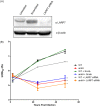
Requirement of LARP7 for intracellular replication of L. pneumophila. (A) Cells were treated with LARP7 RNAi for 24 h then infected. Knockdown of LARP7 was determined by immunoblotting with anti-LARP7 polyclonal antibody. (B) Intracellular growth kinetics of L. pneumophila in hMDMs treated with LARP7-specific or scrambled RNAi. The results are representative of three independent experiments performed in triplicate. Statistical analysis was performed using Student's t test (*, P < 0.05).
FIG 4
FIG 4
The crystal structure of AnkH. (A) AnkH consists of 3 domains: N-terminal ankyrin domain (α1 to α8; red), the cysteine proteinase-like domain (α10 to α17 and β3 to β7; cyan and magenta), and the cap domain (β1 and β2, α9, and α18 to α21; wheat). The inset shows a closeup of putative catalytic triad residues H243, D258, and C324. The HIF hydroxylation sites (N59 and N92) are located within the N-terminal domain and are shown in a sphere representation (blue and red). (B) Primary sequence of ankyrin domain. The length of each ankyrin repeat was determined using the consensus sequence based on statistical analysis of 4,000 ankyrin repeat sequences from the PFAM database as proposed by Mosavi et al. (47). Highlighted (colored) letters correspond to α-helices for each domain. The conserved residues are underlined, and the a-helices are shown as cylinders. (C) Superposition of AnkH with Xanthomonas XopD C470A mutant. The cartoon diagram represents superposition of the AnkH cysteine protease-like domain (residues 163 to 342; orange) and the Xanthomonas XopD C470A mutant (PBD identifier ID 2OIX, residues 336 to 515; green). The three β-strands and two α-helices that form the core of the domains and overlap well are marked. The inset shows a closeup of the catalytic triad. In AnkH, these residues are His243, Asp258, and Cys324; in XopD, these residues are His409, Asp429, and Cys470.
FIG 5
FIG 5
Substitutions in ARDs alter binding efficiency of AnkH and LARP7. (A) The ankyrin domain of AnkH shown as a ribbon diagram. The ankyrin domain consists of four ankyrin repeats: N-cap, repeat 1, repeat2, and C-cap. (B and C) Crystal structure of AnkH illustrating the whole structure (B) and insets representing different locations within the ARDs where residues were substituted (C). (D) Bacterial lysates from WT L. pneumophila and each of the AnkH substitution mutant strains were tested by immunoblotting for AnkH to determine protein stability. Cell lysates were subjected to immunoblotting to detect the presence of AnkH using goat anti-AnkH (53, 56). Equal numbers of bacteria were lysed for each strain. (E) HEK293T cells were transiently transfected with 3XFLAG-AnkH or the indicated 3XFLAG-AnkH substitution mutants and c-myc-LARP7. Densitometry was determined in accordance as actin ratio. (F) Cell lysates were immunoprecipitated with anti-FLAG antibody, and the co-IP was subjected to immunoblotting to detect the presence of AnkH and LARP7. Densitometry of the blotswas determined as the LARP7-to-AnkH ratio.
FIG 6
FIG 6
Structure-function analysis of AnkH in intracellular growth of L. pneumophila within hMDMs. Intracellular growth kinetics were determined for the WT strain, the ankH mutant, or the ankH mutant complemented with the WT allele (c.ankH) or with single and multiple substitution variants as indicated. All strains represented in all the panels were tested using the same WT control. (A) Mutations within first ANK repeat. (B) Mutations within second ANK repeat. (C) Mutations within third ANK repeat. (D) Mutations within the asparagine hydroxylation motif. (E) Mutations within the cystine-like protease pocket. The results are representative of three independent experiments performed in triplicate. Statistical analysis was performed using Student's t test (*, P < 0.05).
FIG 7
FIG 7
Working model of AnkH-LARP7 interaction. In uninfected cells or during ΔankH mutant infection of HEK293T cells, formation of the 7SK snRNP begins when the 5′ methyl capping enzyme (MePCE) and LARP7 are recruited to the 7SK snRNA, forming the core of the 7SK snRNP. After core formation, the HEXIM1/2 dimers as well as the P-TEFb (Cdk9 and cyclin T1) kinase are recruited to complete the 7SK snRNP complex, which prevents transcription elongation by holding RNA polymerase (Pol) II in a paused state. During infection with WT L. pneumophila, AnkH is trafficked to the nucleus, where it interacts with a portion of available LARP7 in the cell. The interaction between AnkH and LARP7 results in a partial inhibition of the 7SK snRNP complex function, leading to enhanced transcriptional elongation by blocking the recruitment of HEXIM1/2 and P-TEFb. The remaining LARP7 present in the cell (the fraction that does not interact with AnkH) is available to interact with other components of the 7SK snRNP complex to pause transcription elongation by preventing P-TEFb from phosphorylating RNA polymerase 2, keeping the polymerase in a paused state. This balance between the pause and relief of the pause in transcriptional elongation results in transcriptional reprogramming within host cell that enhance permissiveness to L. pneumophila infection. There are likely other unidentified substrates of AnkH that could aid in this process or the could act independently of the interaction with LARP7. The effect on amoeba host transcription by AnkH may be different from that seen with human macrophages.

Similar articles

Cited by

References

    1. Abu Khweek A, Amer AO. 2018. Factors mediating environmental biofilm formation by Legionella pneumophila. Front Cell Infect Microbiol 8:38. doi:10.3389/fcimb.2018.00038. - DOI - PMC - PubMed
    1. Fields BS. 1996. The molecular ecology of legionellae. Trends Microbiol 4:286–290. doi:10.1016/0966-842X(96)10041-X. - DOI - PubMed
    1. Horwitz MA, Silverstein SC. 1980. Legionnaires’ disease bacterium (Legionella pneumophila) multiplies intracellularly in human monocytes. J Clin Invest 66:441–450. doi:10.1172/JCI109874. - DOI - PMC - PubMed
    1. Rowbotham TJ. 1980. Preliminary report on the pathogenicity of Legionella pneumophila for freshwater and soil amoebae. J Clin Pathol 33:1179–1183. doi:10.1136/jcp.33.12.1179. - DOI - PMC - PubMed
    1. Swart AL, Harrison CF, Eichinger L, Steinert M, Hilbi H. 2018. Acanthamoeba and dictyostelium as cellular models for Legionella infection. Front Cell Infect Microbiol 8:61. doi:10.3389/fcimb.2018.00061. - DOI - PMC - PubMed

Publication types

MeSH terms