

Lamellar hole-associated epiretinal membrane is a common feature of macular holes in retinitis pigmentosa
- PMID: 31455903
- PMCID: PMC7093403
- DOI: 10.1038/s41433-019-0563-3
Lamellar hole-associated epiretinal membrane is a common feature of macular holes in retinitis pigmentosa
Erratum in
-
Correction: Lamellar hole-associated epiretinal membrane is a common featureof macular holes in retinitis pigmentosa.Eye (Lond). 2020 Apr;34(4):788. doi: 10.1038/s41433-020-0765-8. Eye (Lond). 2020. PMID: 31942028 Free PMC article.
Abstract
Objective: To describe the features and surgical outcomes of macular holes (MHs) in patients with retinitis pigmentosa (RP).
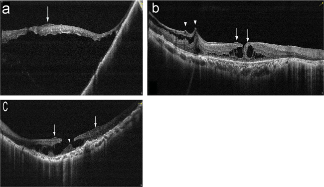
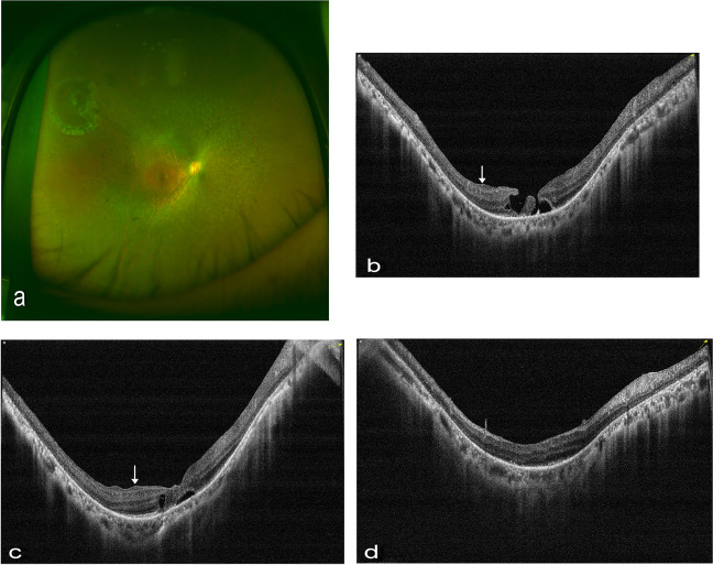
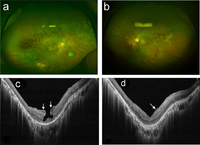
Methods: A review of consecutive series of 110 patients (206 eyes) with RP who underwent comprehensive ophthalmic examinations was conducted. Eleven eyes of ten RP patients were identified with MHs (full thickness or lamellar). Atypical epiretinal membrane, which appeared on spectral-domain optical coherence tomography (SD-OCT) images as a thick homogenous layer of moderately reflective material that was present on the inner retinal layer, was considered to be lamellar hole-associated epiretinal proliferation (LHEP). Five eyes underwent modified vitreoretinal surgery, where hole margin LHEP tissue was retained.
Results: Nine eyes exhibited a lamellar macular hole (LMH), one exhibited a full-thickness macular hole (FTMH), and one exhibited both FTMH and LMH. LHEP was found in all eyes, identified intraoperatively as yellowish, sticky epiretinal membrane with internal limiting membrane beneath it. Two eyes experienced spontaneous closure of MHs without visual acuity (VA) improvement. Five eyes that underwent surgery achieved sealed MHs post-operatively and demonstrated improved, but limited, vision at their latest follow-up.
Conclusions: LHEP is common in MHs associated with RP. While some eyes could achieve spontaneous closure without any VA changes, a conservative vitreoretinal surgery approach, in which the hole margin LHEP tissue is spared, can effectively repair these MHs with limited VA improvement.
Conflict of interest statement
The authors declare that they have no conflict of interest.
Figures
Similar articles
-
Pre- and postoperative OCT features and surgical outcomes of advanced retinitis pigmentosa with macular hole: case series and literature review.BMC Ophthalmol. 2024 Aug 26;24(1):370. doi: 10.1186/s12886-024-03643-y. BMC Ophthalmol. 2024. PMID: 39187836 Free PMC article. Review.
-
Surgical outcomes of lamellar macular holes with and without lamellar hole-associated epiretinal proliferation.Acta Ophthalmol. 2017 May;95(3):e221-e226. doi: 10.1111/aos.13245. Epub 2016 Sep 20. Acta Ophthalmol. 2017. PMID: 27647708
-
Modified surgical technique for lamellar macular holes with lamellar hole-associated epiretinal proliferation (LHEP).Int Ophthalmol. 2021 Jun;41(6):2197-2204. doi: 10.1007/s10792-021-01780-7. Epub 2021 Mar 17. Int Ophthalmol. 2021. PMID: 33730313
-
Epiretinal proliferation seen in association with lamellar macular holes: a distinct clinical entity.Retina. 2014 Aug;34(8):1513-23. doi: 10.1097/IAE.0000000000000163. Retina. 2014. PMID: 24732699
-
[Lamellar macular holes with hyporeflective epiretinal proliferation : OCT diagnostics and clinical course].Ophthalmologe. 2017 Dec;114(12):1100-1109. doi: 10.1007/s00347-017-0597-5. Ophthalmologe. 2017. PMID: 29110126 Review. German.
Cited by
-
Pre- and postoperative OCT features and surgical outcomes of advanced retinitis pigmentosa with macular hole: case series and literature review.BMC Ophthalmol. 2024 Aug 26;24(1):370. doi: 10.1186/s12886-024-03643-y. BMC Ophthalmol. 2024. PMID: 39187836 Free PMC article. Review.
-
UNILATERAL MACULAR HOLE IN A PATIENT WITH RETINITIS PIGMENTOSA TREATED WITH COVER FLAP TECHNIQUE WITH THE USE OF PLATELET-RICH PLASMA UNDER AIR TAMPONADE.Retin Cases Brief Rep. 2025 Jan 1;19(1):84-90. doi: 10.1097/ICB.0000000000001491. Epub 2024 Dec 13. Retin Cases Brief Rep. 2025. PMID: 37756670 Free PMC article.
-
When do patients with retinitis pigmentosa present to ophthalmologists? A multi-centre retrospective study.Eye (Lond). 2024 Dec;38(18):3595-3600. doi: 10.1038/s41433-024-03368-8. Epub 2024 Sep 25. Eye (Lond). 2024. PMID: 39322768
-
Management of Full-Thickness Macular Hole in A Genetically Confirmed Case with Usher Syndrome.Ophthalmol Ther. 2020 Sep;9(3):677-684. doi: 10.1007/s40123-020-00276-4. Epub 2020 Jun 21. Ophthalmol Ther. 2020. PMID: 32566994 Free PMC article.
-
Flap technique-assisted surgeries for advanced retinitis pigmentosa complicated with macular hole: a case report and literature review.BMC Ophthalmol. 2021 Sep 6;21(1):322. doi: 10.1186/s12886-021-02082-3. BMC Ophthalmol. 2021. PMID: 34488687 Free PMC article. Review.
References
-
- Giusti C, Forte R, Vingolo EM. Clinical pathogenesis of macular holes in patients affected by retinitis pigmentosa. Eur Rev Med Pharm Sci. 2002;6:45–8. - PubMed
Publication types
MeSH terms
LinkOut - more resources
Full Text Sources
