Changes in the composition and architecture of staphylococcal biofilm by nisin
- PMID: 31456169
- PMCID: PMC6863270
- DOI: 10.1007/s42770-019-00135-w
Changes in the composition and architecture of staphylococcal biofilm by nisin
Abstract
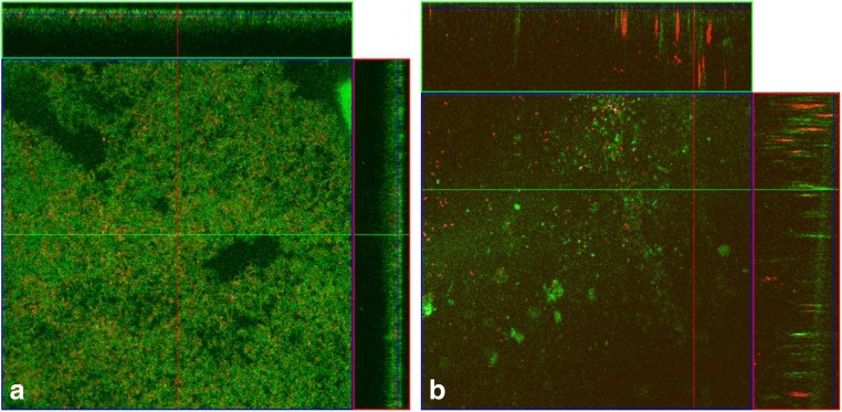
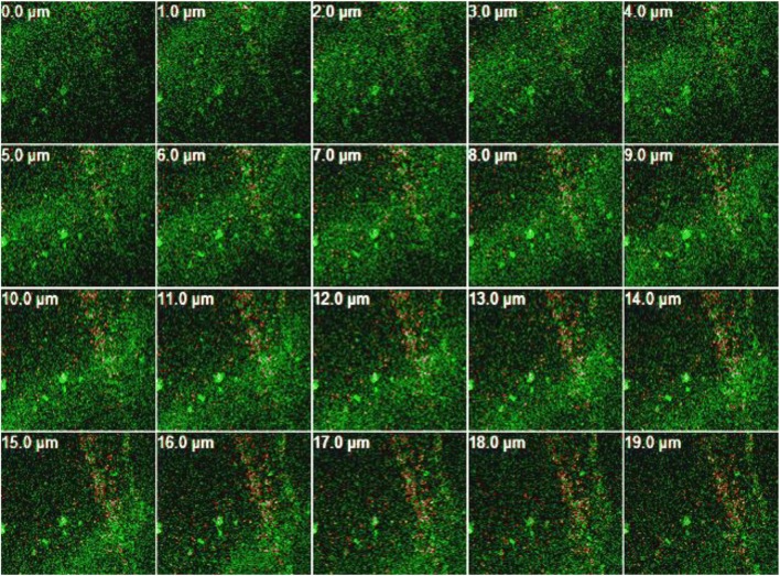
Bacterial biofilms are involved in various medical infections and food contamination episodes and, for this reason, it is of great importance to developing new strategies of its prevention and control. The subinhibitory concentration of nisin was determined, and its effect against Staphylococcus aureus and Staphylococcus epidermidis biofilms was evaluated. Results obtained by confocal laser microscopy demonstrated morphological changes in the architecture of the structure of biofilms. The main components (polysaccharides, proteins, and extracellular DNA (eDNA)) of the biofilm matrix were determined by spectrophotometry and showed that the formation of staphylococcal biofilms in the presence of nisin results in a less dense matrix structure with modification in its constituents. These results contribute to increase the knowledge of the composition and architecture of the extracellular matrix of biofilms of S. aureus, as well as evidence that the investigation of alternative products to assist in the control and combat of biofilms is a promising strategy.
Keywords: Bacteriocin; Biofilm composition; Nisin subinhibitory concentrations; Staphylococcus aureus.
Conflict of interest statement
The authors declare that they have no conflict of interest.
Figures


References
-
- Shale K, Lues JFR, Venter P, Buys EM. The distribution of Staphylococcus sp. on bovine meat from abattoir deboning rooms. Food Microbiol. 2005;22:433–438. doi: 10.1016/j.fm.2004.09.007. - DOI
-
- Di Ciccio P, Vergara A, Festino AR, Paludi D, Zanardi E, Ghidini S, Ianieri A. Biofilm formation by Staphylococcus aureus on food contact surfaces: relationship with temperature and cell surface hydrophobicity. Food Control. 2015;50:930–936. doi: 10.1016/j.foodcont.2014.10.048. - DOI
-
- Abdallah M, Khelissa O, Ibrahim A, Benoliel C, Heliot L, Dhulster P, Chihib NE. Impact of growth temperature and surface type on the resistance of Pseudomonas aeruginosa and Staphylococcus aureus biofilms to disinfectants. Int J Food Microbiol. 2015;214:38–47. doi: 10.1016/j.ijfoodmicro.2015.07.022. - DOI - PubMed
MeSH terms
Substances
Grants and funding
LinkOut - more resources
Full Text Sources
Medical
Molecular Biology Databases