. 2019 Nov;30(11):1371-1384.
doi: 10.1089/hum.2019.159. Epub 2019 Sep 26.

Helper-Dependent Adenovirus Transduces the Human and Rat Retina but Elicits an Inflammatory Reaction When Delivered Subretinally in Rats

Affiliations

Helper-Dependent Adenovirus Transduces the Human and Rat Retina but Elicits an Inflammatory Reaction When Delivered Subretinally in Rats

Ian C Han et al. Hum Gene Ther. 2019 Nov.

Abstract

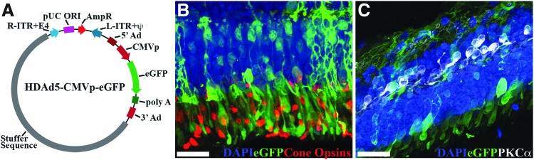
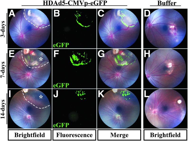
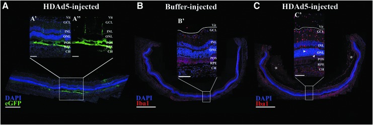
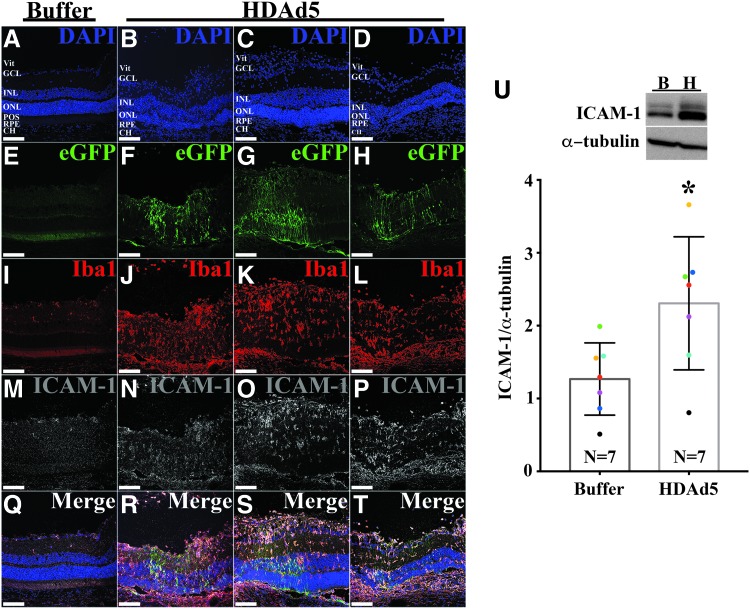
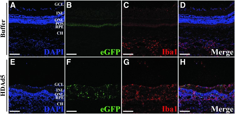
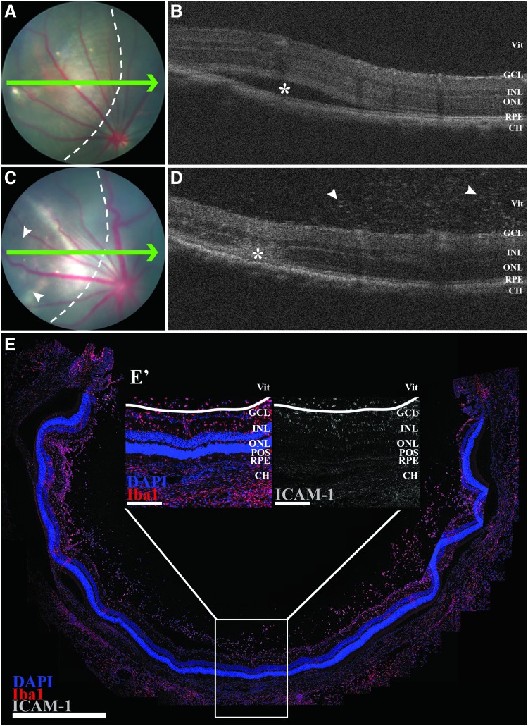
The identification of >100 genes causing inherited retinal degeneration and the promising results of recent gene augmentation trials have led to an increase in the number of studies investigating the preclinical efficacy of viral-mediated gene transfer. Despite success using adeno-associated viruses, many disease-causing genes, such as ABCA4 or USH2A, are too large to fit into these vectors. One option for large gene delivery is the family of integration-deficient helper-dependent adenoviruses (HDAds), which efficiently transduce postmitotic neurons. However, HDAds have been shown in other organ systems to elicit an immune response, and the immunogenicity of HDAds in the retina has not been characterized. In this study, HDAd serotype 5 (HDAd5) was found to successfully transduce rod and cone photoreceptors in ex vivo human retinal organ cultures. The ocular inflammatory response to subretinal injection of the HDAd5 was evaluated using a rat model. Subretinal injection of HDAd5 carrying cytomegalovirus promoter-driven enhanced green fluorescent protein (HDAd5-CMVp-eGFP) elicited a robust inflammatory response by 3 days postinjection. This reaction included vitreous infiltration of ionized calcium-binding adapter molecule 1 (Iba1)-positive monocytes and increased expression of the proinflammatory protein, intercellular adhesion molecule 1 (ICAM-1). By 7 days postinjection, most Iba1-positive infiltrates migrated into the neural retina and ICAM-1 expression was significantly increased compared with buffer-injected control eyes. At 14 days postinjection, Iba1-positive cells persisted in the retinas of HDAd5-injected eyes, and there was thinning of the outer nuclear layer. Subretinal injection of an empty HDAd5 virus was used to confirm that the inflammatory response was in response to the HDAd5 vector and not due to eGFP-induced overexpression cytotoxicity. Subretinal injection of lower doses of HDAd5 dampened the inflammatory response, but also eGFP expression. Despite their larger carrying capacity, further work is needed to elucidate the inflammatory pathways involved and to identify an immunomodulation paradigm sufficient for safe and effective transfer of large genes to the retina using HDAd5.

Keywords: helper-dependent adenovirus; inflammation; retina.


Conflict of interest statement

No competing financial interests exist for any of the authors of this article.

Figures

<b>Figure 1.</b>
Figure 1.
HDAd5-CMVp-eGFP transduces human photoreceptors. (A) Schematic depicting HDAd5-CMVp-eGFP vector plasmid. R-ITR+E4, right-inverted terminal repeat+E4 portion of adenovirus gene; pUC ORI, pUC plasmid origin of replication; AmpR, ampicillin resistance cassette; L-ITR-Ψ, left-inverted terminal repeat+packaging signal; 5′Ad, 5′ adenovirus sequence; CMVp, cytomegalovirus promoter; eGFP, enhanced green fluorescent protein; poly A, polyadenylation; 3′ Ad, 3′ adenovirus sequence. (B, C) Confocal micrographs showing HDAd5-driven eGFP (green) expression in human retinal explants and immunohistochemical labeling with anticone opsins (B; red) and anti-PKCα (C; white) antibodies. Retinal nuclei were counterstained with DAPI (blue). Scale bars = 50 μm. DAPI, 4′,6-diamidino-2-phenylindole dihydrochloride; HDAd, helper-dependent adenoviruses.
<b>Figure 2.</b>
Figure 2.
Subretinal injection of HDAd5-CMVp-eGFP induces atrophic chorioretinal scarring in rat eyes. (A–L) Representative bright field color fundus and eGFP-positive fluorescent images at 3 days (A–C), 7 days (E–G), or 14 days (I–K) postinjection of HDAd5-CMVp-eGFP virus. Representative bright field color fundus images of sucrose buffer-injected contralateral control eyes are also shown at 3 days (D), 7 days (H), and 14 days (L) postinjection. Merged images of color fundus and eGFP fluorescence for HDAd5-injected eyes are shown in (C), (G), and (K). Dotted lines demarcate the boundary of each subretinal injection bleb in (A), (E), and (I). Asterisks in (E) and (I) denote chorioretinal pigmentary atrophic changes. eGFP, enhanced green fluorescent protein.
<b>Figure 3.</b>
Figure 3.
Subretinal injection of HDAd5-CMVp-eGFP induces cellular infiltration into the vitreous by 3 days postinjection. (A) Representative (n = 8; 4 males and 4 females, injected in two independent cohorts) panoramic confocal image depicting site of injection of HDAd5-CMVp-eGFP (eGFP; green) at 3 days postinjection. Transduction and eGFP expression are observed in the RPE and throughout the neural retina (A’, A’’). DAPI (blue) was used to counterstain cell nuclei. (B, C) Panoramic confocal images displaying an entire section through a buffer-injected (B, B’) and an HDAd5-injected (C, C’) eye at 3 days postinjection labeled with Iba1 (red) and the nuclear counterstain, DAPI (blue). White asterisks in (C) denote large clusters of vitreous cells, and white arrowheads (C’) point to the presence of Iba1-positive cells within the outer nuclear layer in a vector-treated eye. The solid white line in each inset demarcates the boundary between the GCL and the vitreous (Vit). Scale bars: (A) 400 μm, (A’, A’’) 100 μm, (B, C) 1000 μm, and (B’ C’) 100 μm. CH, choroid; Iba1, ionized calcium-binding adapter molecule 1; GCL, ganglion cell layer; INL, inner nuclear layer; ONL, outer nuclear layer; POS, photoreceptor outer segments; RPE, retinal pigmented epithelium.
<b>Figure 4.</b>
Figure 4.
HDAd5-induced inflammatory infiltrates express ICAM-1. (A, B) Representative (n = 8; 4 males and 4 females, injected in two independent cohorts) fluorescent confocal micrographs of a buffer- (A) and HDAd5-injected (B) eye demonstrating expression of eGFP (green) and labeled with anti-Iba1 (red) and anti-ICAM-1 (gray). Cell nuclei were counterstained with DAPI (blue). Individual fluorophores are shown below each merged image. Scale bars = 100 μm in each image and definition of abbreviations is same as in Fig. 3. (C) Representative Western blot (n = 7) comparing protein levels of ICAM-1in buffer-injected (lane B) and HDAd5-injected (lane H) whole vitreous/retinal lysates at 3 days postinjection. α-tubulin was used as a loading control. Semiquantitative comparison between buffer- and vector-injected eyes shows a statistically significant increase (*) in ICAM-1 expression (p < 0.05). Like-colored dots represent contralateral eyes from the same animal. ICAM-1, intercellular adhesion molecule 1.
<b>Figure 5.</b>
Figure 5.
By 7 days postinjection of HDAd5, most vitreous cell infiltrates have migrated into the neural retina. (A–T) Representative (n = 10; 5 males and 5 females) confocal fluorescent images comparing a buffer-injected eye (A, E, I, M, Q) to three independent eyes given HDAd5-CMVp-eGFP (B, F, J, N and R; C, G, K, O and S; D, H, L, P, and T) at 7 days postinjection. Transduction of HDAd5 induces expression of eGFP (green; F–H) compared with a buffer-injected control eye (E). Sections are also labeled with anti-Iba1 (red; I–L), anti-ICAM-1 (gray; M–P), and the nuclear counterstain, DAPI (blue; A–D). Merged images for each eye are shown in (Q–T). Definition of abbreviations is same as in Fig. 3. Scale bars = 100 μm in each image. (U) Representative Western blot (n = 7) comparing protein levels of ICAM-1 in buffer-injected (lane B) and HDAd5-injected (lane H) whole vitreous/retinal lysates at 7 days postinjection. α-tubulin was used as a loading control. Semiquantitative comparison between buffer- and vector-injected eyes shows a statistically significant (*) in ICAM-1 expression (p < 0.05). Colored dots represent contralateral eyes from the same animal.
<b>Figure 6.</b>
Figure 6.
The inflammatory response to HDAd5 leads to death of photoreceptors and thinning of the outer nuclear layer. Representative (n = 10; 5 males and 5 females) fluorescent confocal microscopic images of buffer- and HDAd5-injected eyes at 14 days after injection. Sections were counterstained with DAPI to label nuclei (blue; A, E) and labeled with anti-Iba1 (red; C, G). eGFP (green; B, F) was also assessed and only expressed in HDAd5-injected eyes. Merged images for buffer- and HDAd5-injected are shown in (D) and (H), respectively. Definition of abbreviations is same as in Fig. 3. Scale bars = 100 μm in each image.
<b>Figure 7.</b>
Figure 7.
The inflammatory response to HDAd5 is present using an empty vector that does not drive expression of eGFP. Fundus images (A, C) and OCT scans (B, D) immediately after (A, B) and 3 days (C, D) postsubretinal injection of HDAd5E in the same eye. Green arrows in (A) and (C) correspond to location of each OCT line scan. Dotted lines in (A) and (C) denote margin of injection bleb. Asterisk in (B) demarcates subretinal bleb created by injection and asterisk in (D) marks the site of injection after bleb resolution 3 days postinjection. White arrowheads in (D) point to vitreous cell infiltrates. (E) Representative panoramic confocal image displaying an entire section through an HDAd5E-injected (E’) eye at 3 days postinjection. OCT, optical coherence tomography.
<b>Figure 8.</b>
Figure 8.
Decreasing the dose of HDAd5-CMVp-eGFP by 3 log units abrogates inflammatory cellular infiltration, but also yields no visible retinal transduction. (A–C) Representative (n = 3 eyes injected per dose) confocal images at 3 days postinjection near the site of injection of HDAd5-CMVp-eGFP (eGFP; green) at each of three different doses: 5 × 109 vg (A), 5 × 108 vg (B), and 5 × 107 vg (C). Each image is labeled with anti-Iba1 (red), anti-ICAM-1 (gray), and the nuclear counterstain, DAPI (blue). Scale bars = 100 μm. Definition of abbreviations for retinal layers is the same as Fig. 3.

References

    1. Bainbridge JW, Smith AJ, Barker SS, et al. . Effect of gene therapy on visual function in Leber's congenital amaurosis. N Engl J Med 2008;358:2231–2239 - PubMed
    1. Bainbridge JW, Mehat MS, Sundaram V, et al. . Long-term effect of gene therapy on Leber's congenital amaurosis. N Engl J Med 2015;372:1887–1897 - PMC - PubMed
    1. Russell S, Bennett J, Wellman JA, et al. . Efficacy and safety of voretigene neparvovec (AAV2-hRPE65v2) in patients with RPE65-mediated inherited retinal dystrophy: a randomised, controlled, open-label, phase 3 trial. Lancet 2017;390:849–860 - PMC - PubMed
    1. Dimopoulos IS, Hoang SC, Radziwon A, et al. . Two-year results after AAV2-mediated gene therapy for choroideremia: the Alberta experience. Am J Ophthalmol 2018;193:130–142 - PubMed
    1. Cukras C, Wiley HE, Jeffrey BG, et al. . Retinal AAV8-RS1 gene therapy for X-linked retinoschisis: initial findings from a phase I/IIa trial by intravitreal delivery. Mol Ther 2018;26:2282–2294 - PMC - PubMed

Publication types

Substances

LinkOut - more resources