Review
. 2019 Jan;52(1):81-92.
doi: 10.1055/s-0039-1688537. Epub 2019 May 14.

Surgical Management of Lower Extremity Lymphedema: A Comprehensive Review

Affiliations
Review

Surgical Management of Lower Extremity Lymphedema: A Comprehensive Review

Pedro Ciudad et al. Indian J Plast Surg. 2019 Jan.

Abstract

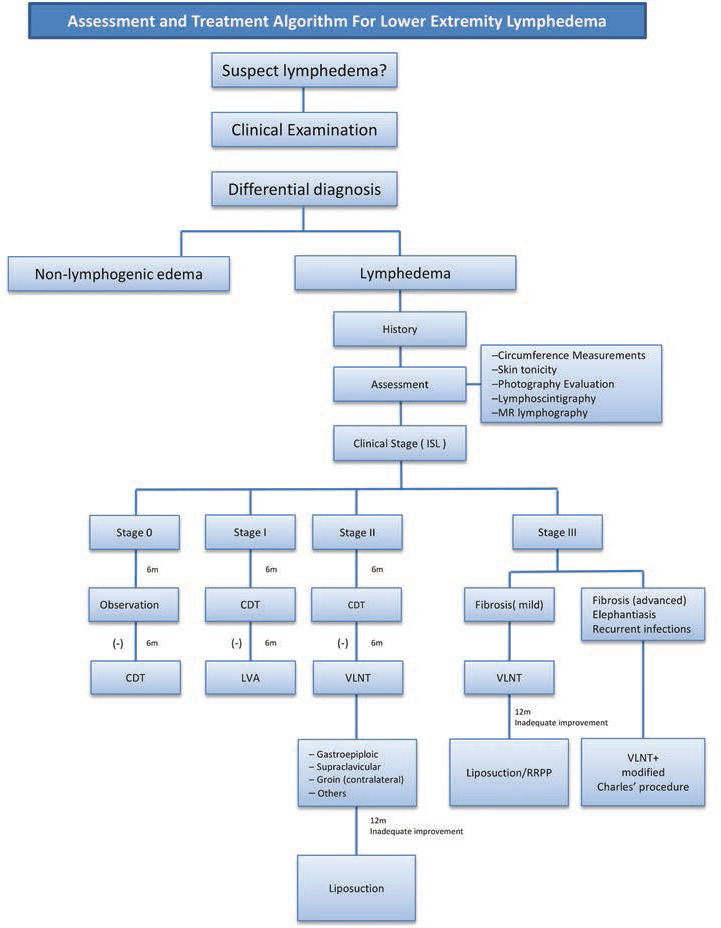
Lymphedema refers to the accumulation of protein-rich fluid in the interstitial spaces. This can occur secondary to congenital malformation of the lymphatic channels or nodes or as a result of an insult that damages appropriately formed channels and nodes. Stagnant, protein-rich lymph initiates an inflammatory response that leads to adipocyte proliferation, fibrous tissue deposition, and increased susceptibility to infections. The end result is permanent disfigurement and dermal changes. Early and accurate diagnosis is essential, since lymphedema is a chronic and progressive problem. When lymphedema affects the lower extremity, it is important to manage it in a way that preserves function and mobility. Early diagnosis also allows for a proactive rather than reactive approach to treatment and utilization of novel physiologic procedures, such as lymphovenous anastomosis and vascularized lymph node transfer. Such interventions slow down disease progression and reduce morbidity by allowing the surgeon to salvage the remaining functional lymphatic channels. When physiologic procedures fail or when faced with a delayed presentation, the addition of excisional procedures can provide a more comprehensive treatment of this debilitating disease. The aim of this article is to review the most current concepts in the surgical management of lower extremity lymphedema.

Keywords: lower extremity lymphedema; lymphedema; radical reduction in lymphedema with preservation of perforators; suction-assisted lipectomy; vascularized lymph node transfer.


Conflict of interest statement

Conflict of Interest None of the authors received any funds or has any financial interests to disclose.

Figures

Fig. 1
Fig. 1
An algorithmic approach is essential for the clinical management of extremity lymphedema. To achieve better outcomes, it helps to optimize the use of diagnostic modalities. For lower extremity lymphedema, the management rests on eliciting detailed history and performing thorough clinical examination. This is effectively complemented by other diagnostic modalities. The options range from simple circumference measurements, measurement of skin tonicity to imaging studies like lymphoscintigraphy and magnetic resonance lymphangiograms. Photographic documentation also plays an important role. The entire evaluation process helps to ascertain whether the etiology is systemic or local, congenital or acquired. Correct diagnosis is the cornerstone in selecting the right procedure for the right patient. The stage of severity can then be decided as per the International Society of Lymphology staging criteria. Stage 0 is the preclinical stage. For these patients, we suggest regular observation for up to 6 months. Depending on whether the swelling subsides or increases, they may be further observed or subjected to compression decongestive therapy (CDT). Stage 1 patients are immediately started on CDT after diagnosis. The decision to intervene is taken 6 months later. The patients who do not respond satisfactorily are ideal candidates for lymphovenous anastomosis (LVA). For stage 2 patients, 6-month trial of CDT may be tried first. The patients with persistent or increasing symptoms vascularized lymph node transfer (VLNT) are offered. The commonly performed vascularized lymph node flaps are gastroepiploic, supraclavicular, and groin. These patients are offered liposuction, if the reduction is not satisfactory at the end of 12 months. For stage 3 patients, CDT trial is not mandatory and immediate intervention is considered. If mild fibrosis is seen, VLNT is performed. Twelve months later (depending on the improvement), one can plan debulking procedure in the form of liposuction or radical reduction in lymphedema with preservation of perforators. However, if the fibrosis is severe along with recurrent infection and features of elephantiasis, debulking with modified Charles’ procedure is the procedure of choice. Ciudad P, Agko M, Chen HC. LYMPHEDEMA - Surgical Approach and Specific Topics. Elsevier Taiwan, Algorithm for the Surgical Management of Lymphedema, 188–189.
Fig. 2
Fig. 2
Patient with left lower extremity lymphedema managed conservatively with complete decongestive therapy (CDT) that encompasses manual lymphatic drainage, compression therapy, and skin/wound care. ( A ) Pre-CDT. ( B ) Three months after CDT.
Fig. 3
Fig. 3
Anastomotic coupler devices for lymphovenous anastomosis.
Fig. 4
Fig. 4
( A ) Anatomical landmarks for intra-abdominal vascularized lymph nodes from the gastroepiploic and ileocecal region. ( B ) The gastroepiploic lymph node flap was based in the right gastroepiploic vessels. The appendicular and the ileocecal vascularized lymph node flaps were based on the appendicular and ileocolic artery and vein, respectively. ( C ) For the vascularized appendicular lymph node flap, appendectomy was performed under the operating microscope on a side table. The mesoappendix was dissected and separated from the appendix while paying attention not to devascularize the flap. ( D ) Vascularized ileocecal lymph node. Both lymph node flaps are alternative options for vascularized lymph node transfer in very selected patients when other more common lymph node flaps are not available.
Fig. 5
Fig. 5
Double level inset by division in half of a single gastroepiploic vascularized lymph node flap is shown. Upper extremity ( A ) and lower extremity ( B ).
Fig. 6
Fig. 6
A double vascularized gastroepiploic lymph node transfer had been performed in one-stage procedure from a single donor site in a patient with bilateral congenital lymphedema.
Fig. 7
Fig. 7
( A ) Preoperative and ( B ) postoperative appearance following radical reduction in lymphedema with preservation of perforators of the left lower extremity.
Fig. 8
Fig. 8
A 46-year-old woman with chronic right lower limb lymphoedema. ( A ) Preoperative appearance showing a right lower which was severely affected by lymphedema. The skin was tensed with multiple ulcers and intermittent leakage of lymph. There were deep skin crypts which collected a lot of sebaceous secretion with foul smell. It was very difficult to remove. The bad smell could be sensed when people stand at a distance away. ( B ) Postoperative picture at 3 years of follow-up after Charles’ procedure.
Fig. 9
Fig. 9
Compressive stocking to prevent hypertrophic scarring after Charles’ procedure.

References

    1. Shimony A, Tidhar D. Lymphedema: a comprehensive review. Ann Plast Surg. 2008;60(02):228. - PubMed
    1. International Society of Lymphology . The diagnosis and treatment of peripheral lymphedema: 2013 Consensus Document of the International Society of Lymphology. Lymphology. 2013;46(01):1–11. - PubMed
    1. Hansen K C, D'Alessandro A, Clement C C, Santambrogio L. Lymph formation, composition and circulation: a proteomics perspective. Int Immunol. 2015;27(05):219–227. - PubMed
    1. Kung T A, Champaneria M C, Maki J H, Neligan P C. Current concepts in the surgical management of lymphedema. Plast Reconstr Surg. 2017;139(04):1003e–1013e. - PubMed
    1. Liao S, von der Weid P Y. Lymphatic syste an active pathway for immune protection. Semin Cell Dev Biol. 2015;38:83–89. - PMC - PubMed