

Review
doi: 10.1055/s-0039-1688536.
Epub 2019 May 17.
Soft Tissue Coverage for Defects around the Knee Joint
Affiliations
- PMID: 31456621
- PMCID: PMC6664846
- DOI: 10.1055/s-0039-1688536
Item in Clipboard
Review
Soft Tissue Coverage for Defects around the Knee Joint
Indian J Plast Surg.
2019 Jan.
Abstract
Soft tissue injuries around the knee present a challenge for providing a cover when there is loss of tissue. Various flaps comprising of skin and muscles around the joint have been described. Understanding the anatomical basis and the design of these flaps can aid in choosing the right flap for a given situation. A prompt cover of the defects aids in quicker healing and quicker rehabilitation of the patient.
Keywords: Knee joint; flap cover; soft tissue defect.
Conflict of interest statement
Figures
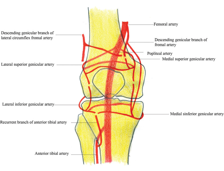
Anastomosis around knee joint.
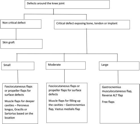
Algorithm for choosing a flap for coverage of knee defects. ALT, anterolateral thigh.
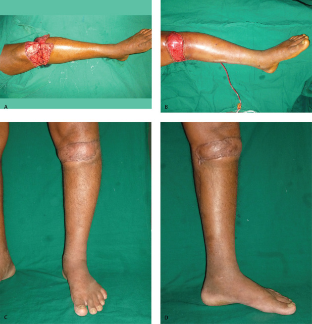
(
A
) Defect around knee joint. (
B
) Gastrocnemius flap covering the defect. (
C
) Postoperative photo—front view. (
D
) Postoperative photo—medial view.
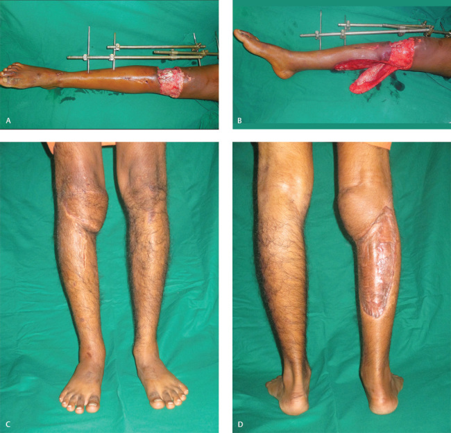
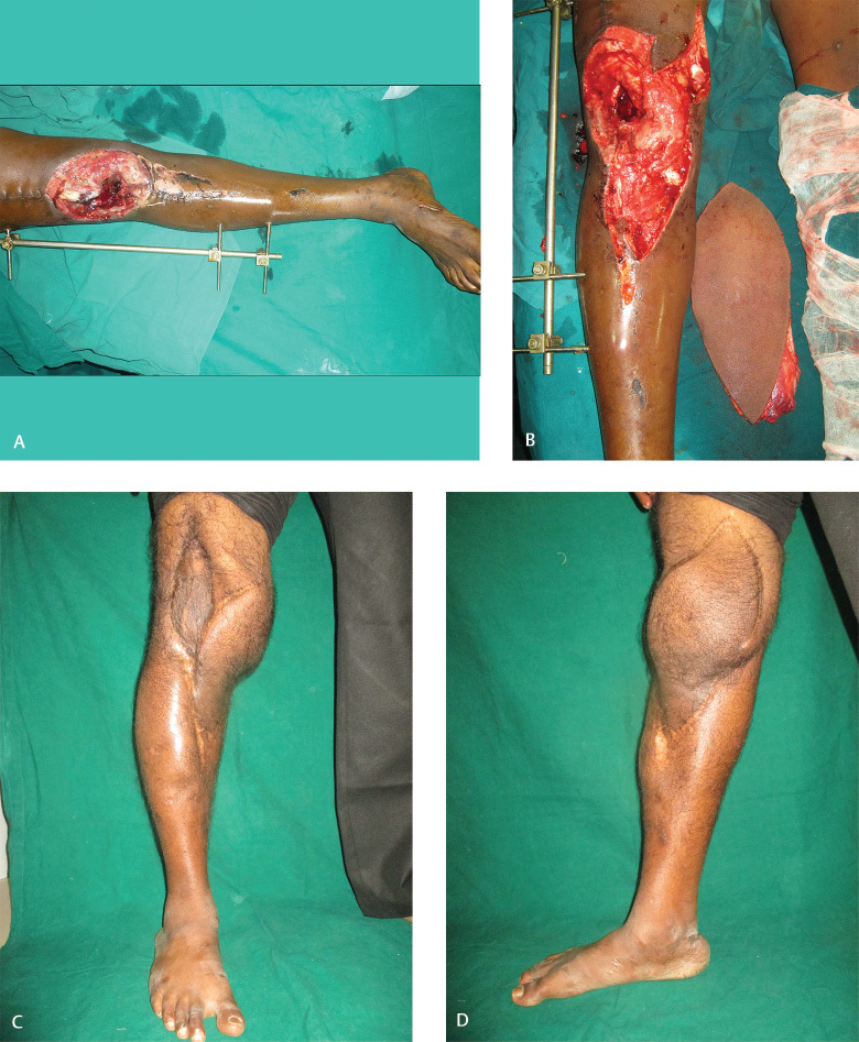
(
A
) Defect over the knee joint. (
B
) Defect with gastrocnemius myocutaneous flap. (
C
) Postoperative photo—front view. (
D
) Postoperative photo—donor site.
(
A
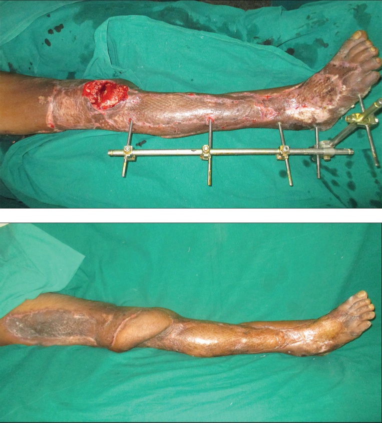
) Defect over knee joint. (
B
) Defect covered with reverse anterolateral thigh flap—postoperative photo.
(
A
) Defect over the knee joint. (
B
) Defect with free anterolateral thigh flap. (
C
) Postoperative photo—front view. (
D
) Postoperative photo—medial view.
References
-
- Salgado C J, Mardini S, Jamali A A, Ortiz J, Gonzales R, Chen H C. Muscle versus nonmuscle flaps in the reconstruction of chronic osteomyelitis defects. Plast Reconstr Surg. 2006;118(06):1401–1411. - PubMed
-
- Cherubino M, Corno M, D'Arpa S.Muscle versus fasciocutaneous flap in lower limb reconstruction: is there a best option? J Reconstr Microsurg 201733(S 01)S27–S33. - PubMed
-
- Cho E H, Shammas R L, Carney M J. Muscle versus fasciocutaneous free flaps in lower extremity traumatic reconstruction: a multicenter outcomes analysis. Plast Reconstr Surg. 2018;141(01):191–199. - PubMed
Publication types
LinkOut - more resources
Full Text Sources