. 2019 Aug 13:13:56.
doi: 10.3389/fncom.2019.00056. eCollection 2019.

Automatic Brain Tumor Segmentation Based on Cascaded Convolutional Neural Networks With Uncertainty Estimation

Affiliations

Automatic Brain Tumor Segmentation Based on Cascaded Convolutional Neural Networks With Uncertainty Estimation

Guotai Wang et al. Front Comput Neurosci. .

Abstract

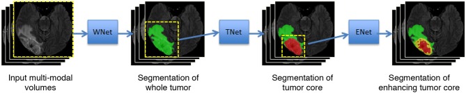
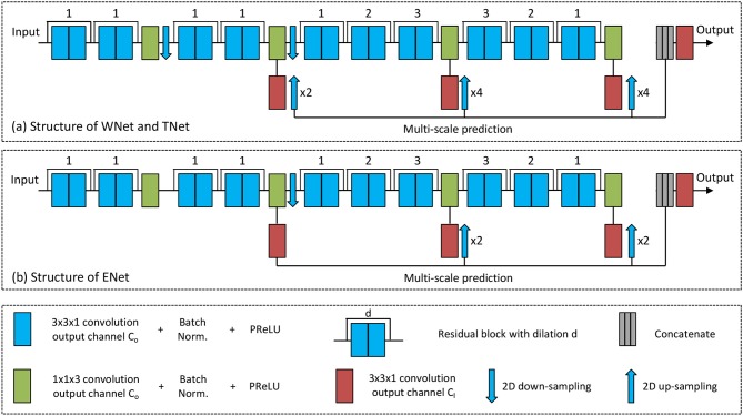
Automatic segmentation of brain tumors from medical images is important for clinical assessment and treatment planning of brain tumors. Recent years have seen an increasing use of convolutional neural networks (CNNs) for this task, but most of them use either 2D networks with relatively low memory requirement while ignoring 3D context, or 3D networks exploiting 3D features while with large memory consumption. In addition, existing methods rarely provide uncertainty information associated with the segmentation result. We propose a cascade of CNNs to segment brain tumors with hierarchical subregions from multi-modal Magnetic Resonance images (MRI), and introduce a 2.5D network that is a trade-off between memory consumption, model complexity and receptive field. In addition, we employ test-time augmentation to achieve improved segmentation accuracy, which also provides voxel-wise and structure-wise uncertainty information of the segmentation result. Experiments with BraTS 2017 dataset showed that our cascaded framework with 2.5D CNNs was one of the top performing methods (second-rank) for the BraTS challenge. We also validated our method with BraTS 2018 dataset and found that test-time augmentation improves brain tumor segmentation accuracy and that the resulting uncertainty information can indicate potential mis-segmentations and help to improve segmentation accuracy.

Keywords: brain tumor segmentation; convolutional neural network; data augmentation; deep learning; uncertainty.


Figures

Figure 1
Figure 1
Our proposed framework with triple cascaded CNNs for multi-modal brain tumor segmentation. We use three CNNs to hierarchically and sequentially segment whole tumor, tumor core and enhancing tumor core, and the CNNs are referred to as WNet, TNet, and ENet, respectively.
Figure 2
Figure 2
The proposed anisotropic CNNs with residual connection, dilated convolution, and multi-scale prediction. Only one downsampling layer is used in ENet as its input size is smaller.
Figure 3
Figure 3
Segmentation results of an HGG brain tumor (A) and an LGG brain tumor (B) from our local validation set, which is part of BraTS 2017/2018 training set. Edema, non-enhancing tumor core and enhancing tumor core are visualized in green, red, and yellow, respectively. White arrows highlight some mis-segmentations.
Figure 4
Figure 4
Examples of test-time augmentation (TTA) combined with different CNNs for brain tumor segmentation. The images are from BraTS 2018 validation set, of which ground truth are not provided by the organizer. In each subfigure, the first row shows the input image of the same patient in four modalities, and the second row shows segmentation results. Edema, non-enhancing tumor core and enhancing tumor core are visualized in green, red, and yellow, respectively. (A,B) Show images of two different patients.
Figure 5
Figure 5
An example of brain tumor segmentation result and the associated voxel-wise uncertainty estimation based on our cascaded CNNs with test-time augmentation (TTA). Taking the uncertainty information for post-processing by conditional random field (CRF) helps to correct the mis-segmented region, as shown in (F). (A) FLAIR, (B) T1ce, (C) Initial segmentation, (D) Voxel-wise uncertainty, (E) Post-process with CRF, (F) Post-process with uncertainty-aware CRF, and (G) Ground truth.
Figure 6
Figure 6
Relationship between segmentation error (1-Dice) and structure-wise uncertainty in terms of volume variation coefficient (VVC) for BraTS 2018 validation set. (A) Enhancing core, (B) Whole tumor, and (C) Tumor core.

References

    1. Abadi M., Barham P., Chen J., Chen Z., Davis A., Dean J., et al. (2016). TensorFlow: A system for large-scale machine learning, in USENIX Symposium on Operating Systems Design and Implementation (Savannah, GA: ), 265–284.
    1. Abdulkadir A., Lienkamp S. S., Brox T., Ronneberger O. (2016). 3D U-Net: Learning dense volumetric segmentation from sparse annotation, in International Conference on Medical Image Computing and Computer Assisted Intervention (Athens: ), 424–432.
    1. Ayhan M. S., Berens P. (2018). Test-time data augmentation for estimation of heteroscedastic aleatoric uncertainty in deep neural networks, in Medical Imaging with Deep Learning (Amsterdam: ), 1–9.
    1. Bakas S., Akbari H., Sotiras A., Bilello M., Rozycki M., Kirby J., et al. (2017a). Segmentation labels and radiomic features for the pre-operative scans of the TCGA-GBM collection. Cancer Imaging Arch. 10.7937/K9/TCIA.2017.KLXWJJ1Q - DOI - PMC - PubMed
    1. Bakas S., Akbari H., Sotiras A., Bilello M., Rozycki M., Kirby J. S., et al. . (2017b). Advancing the cancer genome atlas glioma MRI collections with expert segmentation labels and radiomic features. Nat. Sci. Data 4:170117. 10.1038/sdata.2017.117 - DOI - PMC - PubMed

LinkOut - more resources