. 2019 Aug 13:10:790.
doi: 10.3389/fphys.2019.00790. eCollection 2019.

An Interplay Between Reaction-Diffusion and Cell-Matrix Adhesion Regulates Multiscale Invasion in Early Breast Carcinomatosis

Affiliations

An Interplay Between Reaction-Diffusion and Cell-Matrix Adhesion Regulates Multiscale Invasion in Early Breast Carcinomatosis

Dharma Pally et al. Front Physiol. .

Abstract

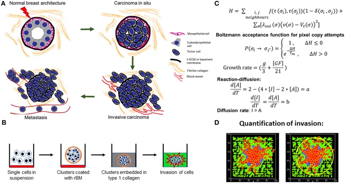
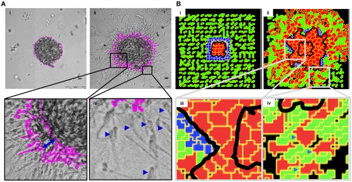
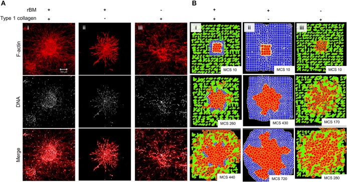
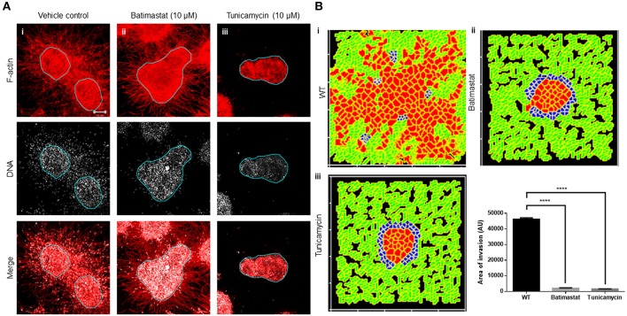
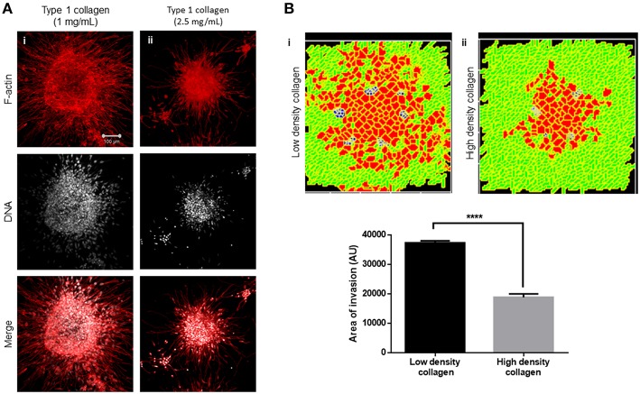
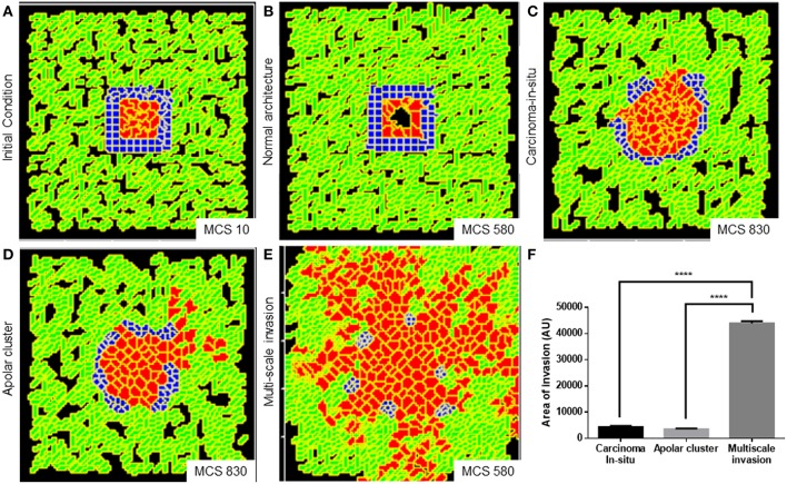
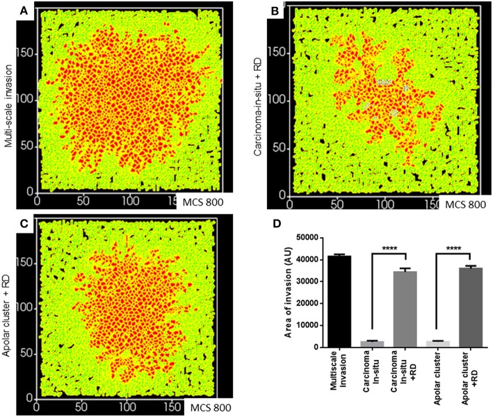
The progression of cancer in the breast involves multiple reciprocal interactions between malignantly transformed epithelia, surrounding untransformed but affected stromal cells, and the extracellular matrix (ECM) that is remodeled during the process. A quantitative understanding of the relative contribution of such interactions to phenotypes associated with cancer cells can be arrived at through the construction of increasingly complex experimental and computational models. Herein, we introduce a multiscale three-dimensional (3D) organo- and pathotypic experimental assay that approximates, to an unprecedented extent, the histopathological complexity of a tumor disseminating into its surrounding stromal milieu via both bulk and solitary motility dynamics. End point and time-lapse microscopic observations of this assay allow us to study the earliest steps of cancer invasion as well as the dynamical interactions between the epithelial and stromal compartments. We then simulate our experimental observations using the modeling environment Compucell3D that is based on the Glazier-Graner-Hogeweg model. The computational model, which comprises adhesion between cancer cells and the matrices, cell proliferation and apoptosis, and matrix remodeling through reaction-diffusion-based morphogen dynamics, is first trained to phenocopy controls run with the experimental model, wherein one or the other matrices have been removed. The trained computational model successfully predicts phenotypes of the experimental counterparts that are subjected to pharmacological treatments (inhibition of N-linked glycosylation and matrix metalloproteinase activity) and scaffold modulation (alteration of collagen density). Further parametric exploration-based simulations suggest that specific permissive regimes of cell-cell and cell-matrix adhesions, operating in the context of a reaction-diffusion-regulated ECM dynamics, promote multiscale invasion of breast cancer cells and determine the extent to which the latter migrate through their surrounding stroma.

Keywords: breast cancer; cell adhesion; cellular potts model (CPM); multiscale invasion; reaction diffusion.


Figures

Figure 1
Figure 1
Schematic depiction of experimental system and computational model. (A) Early stages of breast cancer progression: Top left denotes the normal glandular (ductal/luminal) architecture of human breast. Top right denotes the pattern of breast epithelia undergoing ductal carcinoma in situ where normal epithelia are malignantly transformed, lose polarity, and proliferate, resulting in filling up of ductal lumen. Bottom right shows the architecture of invasive ductal/luminal carcinoma where transformed cells breach the basement membrane (BM) and invade into collagen-rich breast stroma. Bottom left shows how invasive breast cancer cells, having traversed through stroma intravasate into blood/lymph vessels and metastasize to secondary organs. (B) Schematic workflow of 3D invasion assay used throughout the paper: Cells are first cultured on top of nonadhesive substrata in medium containing 4% reconstituted BM (rBM). Once cells assembled into clusters, the latter is embedded within type 1 collagen scaffolds and cultured in serum-free conditions. (C) Governing equations used for setting up the computational model. The first equation calculates H, which is Hamiltonian of the simulation: H determines the probability P associated with index-copy attempts, movements by generalized cells to minimize local effective energy through the default dynamical algorithm known as modified Metropolis dynamics. The equation for calculating growth rate shows it to be dependent on [GF], the growth factor concentration, and g, which denotes nutrient availability. Extracellular matrix (ECM) remodeling follows the kinetics of reaction-diffusion (please refer to the appropriate sections in Material and Methods for a more detailed description of model construction). (D) Quantification of invasiveness of cancer cells in the computational model is performed using the minimal enclosing circle algorithm developed using MATLAB.
Figure 2
Figure 2
Multiscale multicellular invasion of breast cancer cells in culture. (A) Representative phase contrast micrographs from time-lapse imaging of MDA-MB-231 cells showing multiscale invasion into fibrillar matrix. rBM-coated MDA-MB-231 clusters embedded within type 1 collagen (Ai) invade into the latter within 24 h (Aii). Cells show expansive migration [(Aiii); double-headed blue arrow shows the extent of collective migration between initial boundary (0 h) and final boundary (24 h) of the cluster (boundaries shown in pink)]. Single mesenchymal cells are also observed in type 1 collagen [(Aiv) right inset; blue arrowheads]. Scale bar: 200 μm. (B) Multiscale invasion exhibited by computational model. Initial pattern [(Bi); MCS10; cancer cells (red) packed within a BM-like nonfibrillar matrix (blue) and further outward by fibrillar collagen-like matrix (green; inter-fibrillar gap = 3 unit pixels)], and final pattern [(Bii); MCS 440] showing invasion of cells. In silico cells show expansive migration [(Biii) left inset; bulk movement visible through the spatial gap between two black lines denoting boundary at initial MCS and final MCS]. Single cells are also observed in non-fibrillar ECM [(Biv) right inset].
Figure 3
Figure 3
Single-matrix controls of models show simpler modes of invasion. (A) Maximum intensity projections of laser confocal micrographs of MDA-MB-231 cell clusters cultured within specific matrix milieu, fixed and stained for F-actin (using phalloidin; red; top row), DNA (using DAPI; white; middle row), and with both signals merged (bottom row). (i) rBM-coated clusters embedded in type 1 collagen show multiscale invasion (left column). (ii) rBM-coated clusters embedded in rBM show collective or streaming migration of cells, (iii) Uncoated MDA-MB-231 clusters in type 1 collagen show predominantly single cell invasion. Scale bar: 100 μm. (B) Representative images from simulations of invasion of cancer cells at early (top row), intermediate (middle row), and late MCS steps (bottom row). Simulations mimicking cells encapsulated within nonfibrillar and then fibrillar ECM exhibit multiscale invasion (left column). Simulations of cells cultured exclusively in nonfibrillar and fibrillar ECM show collective and single cell migration (Bii and Biii). Interfibrillar gap of C1 = 3 unit pixels.
Figure 4
Figure 4
Inhibition of matrix metalloproteinase activity and N-linked glycosylation inhibit multiscale invasion. (A) Maximum intensity projections of laser confocal micrographs of MDA-MB-231 cell clusters cultured within specific matrix milieu, fixed and stained for F-actin (using phalloidin; red; top row), DNA (using DAPI; white; middle row), and with both signals merged (bottom row). (i) rBM-coated clusters embedded in type 1 collagen treated with vehicle control DMSO show multiscale invasion (left column). (ii) Treatment with 10 μM Batimastat leads to inhibition of transition of cells to type 1 collagen although cytoplasmic projections of cells in the periphery of the cluster are visible in the fibrillar matrix. (iii) Treatment with 10 μM Tunicamycin results in complete abrogation of multiscale invasion. (B) Simulations of control conditions (i), parametric variations analogous with inhibition of R-D (ii), and parametric variations analogous with inhibition of cell–cell, cell–fibrillar ECM, and R-D (iii) at MCS590. Graph represents invasiveness of cells in simulations associated with (Bi–iii). Each bar represents mean ± SEM. ****p < 0.0001.
Figure 5
Figure 5
Increased collagen density impairs multiscale invasion. (A) Maximum intensity projections of laser confocal micrographs of MDA-MB-231 cell clusters cultured within specific matrix milieu, fixed and stained for F-actin (using phalloidin; red; top row), DNA (using DAPI; white; middle row), and with both signals merged (bottom row). (i) rBM-coated clusters embedded in 1 mg/ml type 1 collagen show multiscale invasion. (ii) rBM-coated clusters embedded in 2.5 mg/ml type 1 collagen show impaired invasion of cells into surrounding high-density type 1 collagen. (B) Simulations of control conditions (i) and high-density arrangement of fibrillar ECM showing impaired migration of cells at MCS 540. Graph represents invasiveness of cells in simulations associated with (Bi,ii). Each bar represents mean ± SEM. ****p < 0.0001.
Figure 6
Figure 6
Parameter variation in the computational model can simulate homeostatic, precancerous, and indolently cancerous phenotypes. (A) Initial configuration of all components of the computational model at MCS 0. (B) Simulation of a homeostatic growth-arrested phenotype with a central lumen obtained upon imposing a nonfibrillar ECM-based rules for regulation of cellular quiescence and death of anchored and detached cells, respectively. (C) Simulation of a carcinoma in situ-like phenotype obtained by maintaining high values of cell–cell and cell–nonfibrillar ECM adhesion. (D) Decreasing cell–cell and cell–ECM adhesion in simulation leads to a phenotype that shows further loss of polarity (as evidenced by a roughness in the outer contour of the clusters) and occasional subinvasive single cell phenotypes. (E) Further decreasing cell–cell and cell–matrix adhesion and deployment of an R-D-based kinetics of ECM remodeling leads to multiscale invasion. (F) Quantification (bottom right) of the invasiveness of cells from simulations of homeostasis, carcinoma in situ, apolar clusters, and multiscale invasion. Scale bar: 100 μm. Each bar represents mean ± SEM. ****p < 0.0001.
Figure 7
Figure 7
Simulations of the deployment of R-D-based kinetics in carcinoma in situ and subinvasive cluster phenotypes. (A) Simulation of a control multiscale invasion. (B) Simulation, within which regulation of the ECM was modeled using R-D kinetics in parameter regimes of adhesion cognate to carcinoma in situ phenotypes, predicts collective but not single-cell invasion. (C) Simulations, within which regulation of the ECM was modeled using R-D kinetics in parameter regimes of adhesion cognate to subinvasive apolar cluster phenotypes, predict multiscale invasion. (D) Quantification of the invasiveness of cells from the above simulations in comparison with control multiscale invasion. Each bar represents mean ± SEM. ****p < 0.0001.

Similar articles

Cited by

References

    1. Alber M. S., Kiskowski M. A., Jiang Y. (2004). Two-stage aggregate formation via streams in myxobacteria. Phys. Rev. Lett. 93:068102. 10.1103/PhysRevLett.93.068102 - DOI - PubMed
    1. Bachmeier B. E., Nerlich A. G., Lichtinghagen R., Sommerhoff C. P. (2001). Matrix metalloproteinases (MMPs) in breast cancer cell lines of different tumorigenicity. Anticancer Res. 21, 3821–3828. - PubMed
    1. Balachander G. M., Balaji S. A., Rangarajan A., Chatterjee K. (2015). Enhanced metastatic potential in a 3D tissue scaffold toward a comprehensive in vitro model for breast cancer metastasis. ACS Appl. Mater. Interfaces 7, 27810–27822. 10.1021/acsami.5b09064 - DOI - PubMed
    1. Balduyck M., Zerimech F., Gouyer V., Lemaire R., Hemon B., Grard G., et al. . (2000). Specific expression of matrix metalloproteinases 1, 3, 9 and 13 associated with invasiveness of breast cancer cells in vitro. Clin. Exp. Metastasis 18, 171–178. 10.1023/A:1006762425323 - DOI - PubMed
    1. Bellis S. L. (2004). Variant glycosylation: an underappreciated regulatory mechanism for beta1 integrins. Biochim. Biophys. Acta 1663, 52–60. 10.1016/j.bbamem.2004.03.012 - DOI - PubMed