Review
. 2019 Aug 13:10:1891.
doi: 10.3389/fimmu.2019.01891. eCollection 2019.

Regulation of IgA Production by Intestinal Dendritic Cells and Related Cells

Affiliations
Review

Regulation of IgA Production by Intestinal Dendritic Cells and Related Cells

Hiroyuki Tezuka et al. Front Immunol. .

Abstract

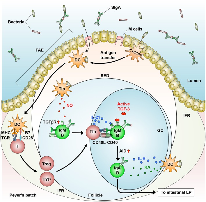
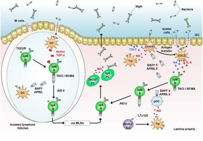
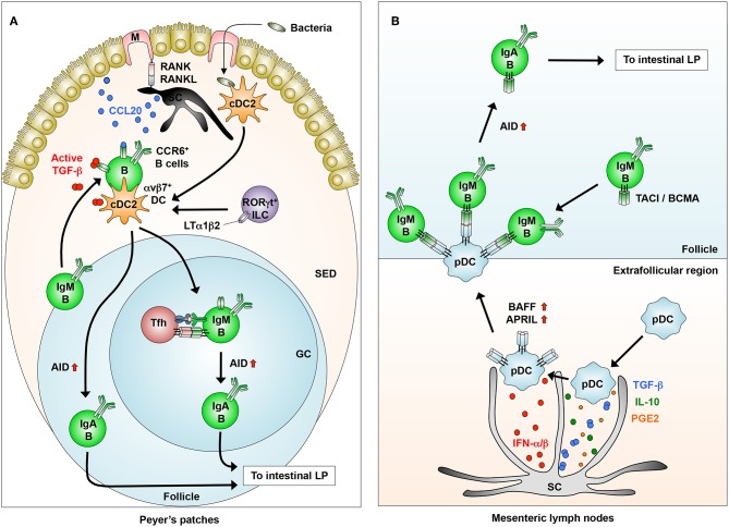
The intestinal mucosa is a physiological barrier for most microbes, including both commensal bacteria and invading pathogens. Under homeostatic conditions, immunoglobulin A (IgA) is the major immunoglobulin isotype in the intestinal mucosa. Microbes stimulate the production of IgA, which controls bacterial translocation and neutralizes bacterial toxins at the intestinal mucosal surface. In the intestinal mucosa, dendritic cells (DCs), specialized antigen-presenting cells, regulate both T-cell-dependent (TD) and -independent (TI) immune responses. The intestinal DCs are a heterogeneous population that includes unique subsets that induce IgA synthesis in B cells. The characteristics of intestinal DCs are strongly influenced by the microenvironment, including the presence of commensal bacterial metabolites and epithelial cell-derived soluble factors. In this review, we summarize the ontogeny, classification, and function of intestinal DCs and how the intestinal microenvironment conditions DCs and their precursors to become the mucosal phenotype, in particular to regulate IgA production, after they arrive at the intestine. Understanding the mechanism of IgA synthesis could provide insights for designing effective mucosal vaccines.

Keywords: IgA; commensal bacteria; conditioning; dendritic cells; intestine.


Figures

Figure 1
Figure 1
T cell-dependent generation of IgA+ B cells in the GALT. In the PPs, dendritic cells (DCs) that engulf directly or indirectly (via antigen transfer by CX3CR1+ cells) luminal bacteria produce IL-6 and move from the SED to the IFR, where they prime CD4+ T cells to generate follicular helper T (Tfh) cells, which are derived from Tregs and Th17 cells. Tfh cells move into the follicle, where they interact with IgM+ B cells in a cognate manner (MHC-TCR and CD40-CD40L). In addition, Tip-DCs induce the expression of TGF-β receptor (TGFβR) through their production of nitric oxide (NO). Subsequently, B cells differentiate into IgA+ B cells through AID expression in response to TGF-β, IL-21 (produced by Tfh cells), and RA (produced by DCs). IgA+ B cells migrate into the intestinal lamina propria (LP), where they differentiate into IgA-producing plasma cells.
Figure 2
Figure 2
T cell-independent generation of IgA+ B cells in the GALT. In the isolated lymphoid follicles, dendritic cells (DCs) that sample luminal bacteria produce TGF-β. DCs also produce BAFF and APRIL to generate IgA+ B cells that home into the lamina propria (LP) and differentiate into IgA-producing plasma cells (PCs). In the LP, CD103+ cDC2 that sample luminal bacteria produce IL-6 and retinoic acid (RA). CX3CR1+ cells produce BAFF and APRIL in response to nitric oxide (NO, produced by Tip-DCs and PCs) and TSLP [produced by intestinal epithelial cells (iECs)]. RORγt+ innate lymphoid cells (ILCs) induce Tip-DCs in an LTα1β2-dependent manner, which produce BAFF and APRIL by pDCs. In the presence of BAFF/APRIL, RA, and IL-6, IgM+ B cells generate IgA+ B cells, which differentiate into IgA-producing PCs.
Figure 3
Figure 3
Novel DC-B cell interaction in inducing IgA synthesis. (A) In the Peyer's patches, stromal cells (SCs) interact with M cells to produce CCL20, which induces the migration of IgM+CCR6+ B cells from the follicles into the SED through the CCR6-CCL20 interaction. B cells that migrated into the SED interact with dendritic cells (DCs) that express integrin αvβ8, which is induced by LTα1β2 from RORγt+ innate lymphoid cells (ILCs). B cells activated in the SED migrate into the follicles to generate IgA+ B cells through the induction of AID. (B) In the extrafollicular region of the mesenteric lymph nodes, SCs condition pDCs to express membrane-bound BAFF/APRIL through their production of IFN-α/β, albeit at low levels, and possibly TGF-β, IL-10, and PGE2. Conditioned pDCs make close contact with IgM+ B cells through BAFF/APRIL-TACI/BCMA interaction to induce AID expression for IgA class-switching.

References

    1. Peterson LW, Artis D. Intestinal epithelial cells: regulation of barrier function and immune homeostasis. Nat Rev Immunol. (2014) 14:141–53. 10.1038/nri3608 - DOI - PubMed
    1. Macpherson AJ, Harris N. Interaction between commneasal intestinal bacteria and the immune system. Nat Rev Immunol. (2004) 4:478–85. 10.1038/nri1373 - DOI - PubMed
    1. Cerutti A. The regulation of IgA class switching. Nat Rev Immunol. (2008) 8:421–34. 10.1038/nri2322 - DOI - PMC - PubMed
    1. Fagarasan S, Kawamoto S, Kanagawa O, Suzuki K. Adaptive Immune regulation in the gut: T cell-dependent and T cell-independent IgA synthesis. Annu Rev Immunol. (2010) 28:243–73. 10.1146/annurev-immunol-030409-101314 - DOI - PubMed
    1. Bunker JJ, Flynn TM, Koval JC, Shaw DG, Meisel M, McDonald BD, et al. . Innate and adaptive humoral responses coat distinct commensal bacteria with immunoglobulin A. Immunity. (2015) 43:541–53. 10.1016/j.immuni.2015.08.007 - DOI - PMC - PubMed

Publication types

Substances