. 2017 Jul:2017:857-860.
doi: 10.1109/ICCA.2017.8003172. Epub 2017 Aug 8.

Pneumatic delivery of untethered microgrippers for minimally invasive biopsy

Affiliations

Pneumatic delivery of untethered microgrippers for minimally invasive biopsy

Andrew Choi et al. IEEE Int Conf Control Autom. 2017 Jul.

Abstract

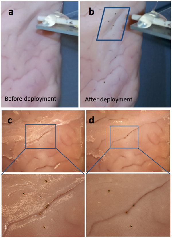
The surgical biopsy is one of the most widely utilized medical procedures for diagnosis of a number of diseases. In order to enable less invasive biopsies, we have previously developed and applied residual stress and physiologically activated sub-millimeter sized untethered grippers. Here, we report a controlled, pneumatic system and methodology for pressurized delivery of untethered microgrippers (μ-grippers) to improve the efficacy of tissue excision. The approach is compatible with current minimally invasive laparoscopic and endoscopic methods. Using a model experimental system, we observed that pneumatic delivery significantly improves the efficiency of the tissue attachment-μ-grippers attach up to 30-fold better on vertically oriented tissues, and up to 3.5-fold better on horizontally oriented tissues as compared to experiments without pressurized delivery. Hence, the use of pneumatics in the delivery of untethered microdevices could significantly enhance their efficiency in minimally invasive biopsy procedures.


Figures

Fig. 1.
Fig. 1.
Optical images of the thermo-sensitive μ-grippers. (A) Time-lapse images of a μ-gripper closing in water at 37°C. (B) Optical image of a μ-gripper closing and gripping three glass beads. (C) Optical image of a μ-gripper closed on pig stomach tissue, ex-vivo. The scale bars represent 200 μm.
Fig. 2.
Fig. 2.. Schematic representation of the pneumatic μ-gripper delivery approach.
Pressurized gas is used to send a bolus of liquid containing μ-grippers onto the tissue.
Fig. 3.
Fig. 3.. Average viability of the μ-grippers after pneumatic release using our integrated system at different pressures.
Viability is defined as the percentage of the μ-grippers that are unbroken after exit from the tube.
Fig. 4.
Fig. 4.. Optical images showing pneumatic dispersal of μ-grippers on tissue in two configurations.
(a) Image of set up with pneumatic dispensation unit and delivery in (b) horizontal and (c) vertical configurations.
Fig. 5.
Fig. 5.
Optical images of μ-grippers on tissue after pneumatic delivery at two pressures (a, b) 0 and (c, d) 3 psi; (a, c) before and (b, d) after flow.
Fig. 6.
Fig. 6.. Graph of attachment percentage vs pressure used for pneumatic delivery on horizontally oriented tissue.
Results show a significant increase in attachment percentage with increasing pressure.
Fig. 7.
Fig. 7.. Optical images of the μ-grippers pneumatically dispensed on vertically oriented tissue.
(a-b) Optical image showing the tissue target before and after μ-gripper pressurized dispensation at 8 psi. (c-d) Progressive zoomed in image of the sample before and after flow showing that some μ-grippers were dislodged after flow.
Fig. 8.
Fig. 8.. Graph of the attachment percentage vs pressure used for pneumatic delivery on vertically oriented tissue.
The results indicate a statistically significant increase in the attachment percentage at pressures of 3 and 8 psi as compared to no pressure.

References

    1. Fernandes R, Gracias DH. “Toward a miniaturized mechanical surgeon,” Mater. Today vol. 12, no. 10, pp: 14–20, 2009
    1. Nelson BJ, Kaliakatsos IK, Abbott JJ. “Microrobots for minimally invasive medicine,” Ann. Rev. Biomed. Eng Vol. 12, pp: 55–85, 2010 - PubMed
    1. Byun SW, et al., “Novel Barbed Micro-Spikes for Micro-Scale Biopsy,” J. Micromech. Microeng, vol. 15, no. 6, pp. 1279–1284, 2005
    1. Gultepe E, Randhawa J, Kadam S, Yamanaka S, Selaru FM, Shin EJ, Kalloo AN, Gracias DH, “Biopsy with Thermally-Responsive Untethered Microtools,” Adv. Mater 25(4), pp.514–519, 2013 - PMC - PubMed
    1. Gultepe E, Yamanaka S, Laflin KE, Kadam S, Shim Y, Olaru AV, Khashab MA, Kalloo AN, Gracias DH, Selaru FM, “Biologic tissue sampling with untethered microgrippers,” Gastroenterology, 144(4), p.691, 2013 - PMC - PubMed

LinkOut - more resources