. 2019 Aug 21;5(2):vez034.
doi: 10.1093/ve/vez034. eCollection 2019 Jul.

Conserved structural RNA domains in regions coding for cleavage site motifs in hemagglutinin genes of influenza viruses

Affiliations

Conserved structural RNA domains in regions coding for cleavage site motifs in hemagglutinin genes of influenza viruses

Alexander P Gultyaev et al. Virus Evol. .

Abstract

The acquisition of a multibasic cleavage site (MBCS) in the hemagglutinin (HA) glycoprotein is the main determinant of the conversion of low pathogenic avian influenza viruses into highly pathogenic strains, facilitating HA cleavage and virus replication in a broader range of host cells. In nature, substitutions or insertions in HA RNA genomic segments that code for multiple basic amino acids have been observed only in the HA genes of two out of sixteen subtypes circulating in birds, H5 and H7. Given the compatibility of MBCS motifs with HA proteins of numerous subtypes, this selectivity was hypothesized to be determined by the existence of specific motifs in HA RNA, in particular structured domains. In H5 and H7 HA RNAs, predictions of such domains have yielded alternative conserved stem-loop structures with the cleavage site codons in the hairpin loops. Here, potential RNA secondary structures were analyzed in the cleavage site regions of HA segments of influenza viruses of different types and subtypes. H5- and H7-like stem-loop structures were found in all known influenza A virus subtypes and in influenza B and C viruses with homology modeling. Nucleotide covariations supported this conservation to be determined by RNA structural constraints that are stronger in the domain-closing bottom stems as compared to apical parts. The structured character of this region in (sub-)types other than H5 and H7 indicates its functional importance beyond the ability to evolve toward an MBCS responsible for a highly pathogenic phenotype.

Keywords: RNA structure; highly pathogenic avian influenza; influenza virus.


Figures

Figure 1.
Figure 1.
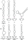
RNA secondary structures encompassing the cleavage site regions of H5 and H7 HA segments, derived from comparative RNA structure predictions (Gultyaev et al. 2016). (A) Alignment of structures based on encoded amino acid sequences at the boundary between HA1 and HA2 regions. The structures are shown using a bracket view with each base pair denoted with a bracket pair. Paired nucleotides are shown with background colors specific for alternative structures. The hairpin loop nucleotides are in bold type and underlined. The stem-loop (SL) structure denoted here as SLH5a is conserved in majority of H5 lineages, while the SLH5b and SLH5c are folded in the lineage of H5N2 viruses first isolated during an outbreak in Mexico. The SLH7a structures are possible in all Eurasian and American H7 strains, the alternative SLH7b folds are conserved only in H7 HA segments of American origin. A small hairpin SLH7c is conserved in H7, H10 and H15 HA RNAs (Gultyaev et al. 2016). Insertions of additional basic amino acid codons in the HPAI strains are shown in red. The N-terminal HA2 glycine amino acid residue is denoted by an asterisk. (B) Examples of conserved structures in LPAI and HPAI viruses. Adapted from Gultyaev et al. (2016), available under the Creative Commons Attribution 4.0 International License
Figure 2.
Figure 2.
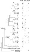
Identification of strains with possible H5- and H7-like domains in different clades of influenza viruses. The ‘−’and ‘+’ signs define predictions of stable structures in at least some or all strains of a given clade, respectively. The prediction of SLH5a-like domains in H9 HA subtype is shown as ‘+/−’ because this structure is not predicted only in a few outlier sequences whereas it is conserved in the overwhelming majority of H9 HA segments. Representative structures are shown in Figs 3–5 and Supplementary Figs S1–S4. The tree topology is according to phylogenetic reconstructions by Russell et al. (2004), Fouchier et al. (2005), Suzuki and Nei (2002), Tong et al. (2013), and Shi et al. (2018). The branches corresponding to geographic sublineages of avian influenza A viruses (Eur., Eurasia; Amer., America), human and swine strains are shown only in cases of different extent of conservation of structures.
Figure 3.
Figure 3.
Alternative rod-like and Y-shaped SLH5a- and SLH7a-like structures in H9 HA RNAs. Covaried nucleotides in the SLH5a domain-closing stems are shown in magenta. The typical SLH5a and SLH7a structures (Gultyaev et al. 2016) are shown for comparison. The GGN codon coding for the N-terminal HA2 glycine amino acid residue is shown by an asterisk. Accessions: A/Duck/Hong Kong/Y280/97(H9N2), AF156376; A/chicken/Sichuan/G2/2009(H9N2), GU471800; A/gadwall/Netherlands/1/2006(H9N2), CY043864. Nucleotide positions of database sequences are given and in case of partial HA sequences the positions corresponding to a full-length segment are shown in brackets. aClades of H9 HA segments are according to Dalby and Iqbal (2014).
Figure 4.
Figure 4.
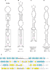
SLH5a-like structures in the influenza B (B) and Wuhan spiny eel influenza (C) viruses. The typical SLH5a structure (A) (Gultyaev et al. 2016) is shown for comparison. Homologous parts of the stems are in dashed frames, covariations are shown in magenta. (D) An alternative stem-loop structure predicted in the HA RNA of Wuhan asiatic toad influenza virus. The folding free energies of structures in fish and amphibian influenza viruses (C, D) are computed at 25 °C in addition to standard values at 37 °C, in order to get approximate values consistent with the natural replication conditions. (E) Alignment of structures. Other notations are similar to Fig. 1. Accessions: B/Lee/40, K00423; Wuhan spiny eel influenza virus, MG600041; Wuhan Asiatic toad influenza virus, MG600048. Only partial sequences of the HA segments of the last two viruses are available.
Figure 5.
Figure 5.
An example of SLH7b-like folding in the influenza C viruses, strain C/Mississippi/80. The SLH7b structures predicted in the HPAI strain A/chicken/Tennessee/17-007147-2/2017(H7N9) and its LPAI progenitor A/chicken/Tennessee/17-007431-3/2017(H7N9) (Lee et al. 2017b) are shown for comparison. Proline codons used as anchors in the HEF1/HA1 alignment are labeled in green. Other notations are similar to Fig. 4.
Figure 6.
Figure 6.
A model of influenza virus polymerase slowdown induced by domain-closing stems in template RNA (vRNA or cRNA). The schematic configuration of entry and exit channels of template RNA, NTP entry, and nascent RNA exit channels is according to modeling by te Velthuis and Fodor (2016).

Similar articles

Cited by

References

    1. Abdelwhab E. M., Veits J., Mettenleiter T. (2013) ‘Genetic Changes That Accompanied Shifts of Low Pathogenic Avian Influenza Viruses toward Higher Pathogenicity in Poultry’, Virulence, 4: 441–52. - PMC - PubMed
    1. Abolnik C. (2017) ‘Evolution of H5 Highly Pathogenic Avian Influenza: Sequence Data Indicate Stepwise Changes in the Cleavage Site’, Archives of Virology, 162: 2219–30. - PubMed
    1. Alexander D. J. (2007) ‘An Overview of the Epidemiology of Avian Influenza’, Vaccine, 25: 5637–44. - PubMed
    1. Bao Y. et al. (2008) ‘The Influenza Virus Resource at the National Center for Biotechnology Information’, Journal of Virology, 82: 596–601. - PMC - PubMed
    1. Canale A. S. et al. (2018) ‘Synonymous Mutations at the Beginning of the Influenza A Virus Hemagglutinin Gene Impact Experimental Fitness’, Journal of Molecular Biology, 430: 1098–115. - PubMed