Review
. 2019 Aug 13:9:761.
doi: 10.3389/fonc.2019.00761. eCollection 2019.

Proteasomes and Several Aspects of Their Heterogeneity Relevant to Cancer

Affiliations
Review

Proteasomes and Several Aspects of Their Heterogeneity Relevant to Cancer

Alexey V Morozov et al. Front Oncol. .

Abstract

The life of every organism is dependent on the fine-tuned mechanisms of protein synthesis and breakdown. The degradation of most intracellular proteins is performed by the ubiquitin proteasome system (UPS). Proteasomes are central elements of the UPS and represent large multisubunit protein complexes directly responsible for the protein degradation. Accumulating data indicate that there is an intriguing diversity of cellular proteasomes. Different proteasome forms, containing different subunits and attached regulators have been described. In addition, proteasomes specific for a particular tissue were identified. Cancer cells are highly dependent on the proper functioning of the UPS in general, and proteasomes in particular. At the same time, the information regarding the role of different proteasome forms in cancer is limited. This review describes the functional and structural heterogeneity of proteasomes, their association with cancer as well as several established and novel proteasome-directed therapeutic strategies.

Keywords: cancer; constitutive proteasome; immunoproteasome; intermediate proteasome; proteasome regulators; spermatoproteasome; thymoproteasome; ubiquitin-proteasome system.


Figures

Figure 1
Figure 1
The structural diversity of proteasomes. There are several levels of proteasome organization (3). Proteasomes differ by composition of subunits forming 20S core particles. Major 20S proteasomes include: constitutive proteasomes, intermediate proteasomes of type I and II, immunoproteasomes. The constitutive 20S proteasomes contain constitutive catalytic subunits: β1, β2, and β5. These proteasomes are found both in the nucleus and the cytoplasm. Up to 85% of all cellular proteasomes contain constitutive 20S cores (3, 5, 14, 20, 21). Thus, constitutive 20S proteasome appears to be the most abundant form of 20S core particle found in various tissues including heart, kidney, skeletal muscles, brain as well as in different cell lines of embryonal or cancer origin (14, 20, 21). 20S immunoproteasome contain immune subunits β1i (LMP2), β2i (MECL-1), and β5i (LMP7) instead of constitutive β1, β2, β5, correspondingly. These complexes were also found both in the nucleus and the cytoplasm (22). Normally, immunoproteasomes are predominantly detected (up to 64% of total proteasome pool) in cells of the immune system as well as in the small bowel and colon, however can be upregulated in many other cells following exposure to the immune cytokines or in stress conditions (3, 14, 23, 24). The intermediate proteasomes contain the immune catalytic subunits together with the constitutive ones. The subcellular localization of intermediate proteasomes was not carefully addressed, and these complexes may be present both in the nucleus and the cytoplasm. The type I (β5i) intermediate proteasomes contain β1, β2, and β5i subunits. In the cellular proteasome pool type I intermediate proteasomes constitute from almost none to more than 50% depending on the tissue and cell type (5, 14). These proteasomes are found in large numbers in the liver, small intestine, colon, muscles (12, 14, 25), dendritic cells (14), as well as in the cells of acute promyelocytic leukemia NB4 and histiocytic lymphoma U937 cell lines (5). The type II intermediate proteasomes contain β1i/β2/β5i catalytic subunits and are abundantly found in monocytes representing up to 54% of the proteasome pool (14). In addition, these proteasomes were detected in several cancer cell lines including acute myelogenous leukemia line KG1a (3, 5). Experimental data indicate that other forms of intermediate proteasomes can be formed e.g., complexes with β1/β2i/β5i (12), β1i/β2i/β5 (26), β1i/β2/β5 (26). Some of them were detected in unnatural conditions (in β5i−/− mice, in cells with β5 overexpression etc.), thus their presence in vivo is debated. Moreover, some of these forms are against rules of cooperative assembly (–29). Nevertheless, their presence in certain situations cannot be entirely excluded. Another level of 20S complex diversity can arise from the fact that each 20S proteasome contain two copies of every subunit imbedded in different rings thus, theoretically, asymmetric proteasomes containing one immune and one constitutive beta-subunit in the same complex can be formed (30, 31). Still this could be a temperate occasional event in the process of proteasome pool reorganization following the stimulation with cytokines. Another level of cellular proteasome organization is a presence of an activator. By several authors it was demonstrated that the majority (depending on the tissue or cell type from 41 to 74%) of cellular proteasomes are “free” and do not bare an activator (5, 13). The activator that is most frequently attached to the 20S proteasomes is the 19S complex. It was found bound to from ~21–57% of 20S complexes in various cells and tissues (5, 13, 32). Proteasomes with 19S activator were detected in the cytoplasm and the nucleus. The 11Sαβ is a second most frequent cytoplasmic proteasome activator and from a single per cent to 44% of cellular proteasomes may be capped with it (5, 13). PA200 and 11Sγ are the regulators, that associate with proteasomes preferentially in the nucleus covering generally up to 8% and <5% of total proteasome pool, respectively (5). PI31 proteasome inhibitor associates with ~1% of cellular 20S proteasomes (5). Whether VCP associates with 20S core particle in mammalian cells and how frequently, is not entirely clear. Interestingly, preferential association of proteasome activators with different 20S core proteasomes was recently shown (15). It was demonstrated that 11Sαβ and 11Sγ “prefer” immune and likely intermediate 20S proteasomes, while PA200 and PI31—the constitutive ones, 19S binds all the complexes equally (15). At the same time, several reports argue preferential association of certain activators with constitutive or immune 20S proteasome (33). The heterogeneity of cellular proteasomes is even greater and the image does not include various post-translational modifications of proteasomes (7) as well as proteasome interacting proteins with regulatory functions (3). Question mark indicates that the presence of a particular proteasome form is uncertain.
Figure 2
Figure 2
Tissue specific proteasomes. Thymoproteasomes contain immune subunits β1i and β2i and a specific catalytic subunit β5t (113). These proteasomes are found exclusively in the thymus and can represent up to 20% of total proteasome pool there, concentrating specifically in cortical thymic epithelial cells where tP is the dominate form of the proteasome (113). Another unique 20S proteasome form is found in the testis. These proteasomes contain α4s subunit together with constitutive, immune or, probably, catalytic β-subunits of both types. Proteasomes with α4s and immune catalytic subunits were named spermatoproteasomes (114). The proportion of proteasomes containing α4s to proteasomes with constitutive α4 subunit in sperm could be as high as 80% (115). Question mark indicates that the presence of a particular proteasome form is uncertain.
Figure 3
Figure 3
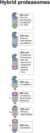
Hybrid proteasomes. These proteasomes represent complexes of two different activators attached to a single 20S particle. Initially hybrids with 19S-20S-11Sαβ and 19S-20S-11Sγ architecture were identified (278). In certain cells 19S-20S-11Sαβ can represent up to 24% of all proteasomes (13). 19S-20S-11Sγ complexes are less frequent (278). 19S-20S-11Sαβ proteasomes are localized in the cytoplasm whereas 19S-20S-11Sγ–in the nucleus. The 20S core particle in 19S-20S-11Sαβ complex is likely immune or intermediate, this is confirmed by the increase of such complexes following IFN-γ treatment (13, 278), still cases with constitutive 20S could not be ruled out (13). 19S-20S-11Sγ could also contain constitutive 20S particle. Another form of hybrid proteasome is the 19S-20S-PA200 (232). The 19S-20S-PA200 proteasomes have nuclear localization, the 20S core particle in these hybrid complexes could be constitutive, or contain α4s subunits (114). Theoretically other hybrid proteasomes: 19S-20S-PI31, PA200-20S-PI31, PA200-20S-11Sγ, and 11Sαβ-20S-PI31 can also exist, although were not revealed so far. Question mark indicates that the presence of a particular proteasome form is uncertain.
Figure 4
Figure 4
Several proteasome-based strategies for cancer therapy. (A) Broad specificity proteasome inhibitors for instance bortezomib affect all forms of proteasomes, thus influencing entire proteolysis in different cells (98). Along these lines, bortezomib is known for strong side-effects that limit its clinical use. To decrease the side-effects and increase the efficacy combinations of bortezomib with other molecules were proposed (283, 284). (B) Subunit specific inhibitors allowed targeting specific subsets of proteasomes and especially iP subunit-specific inhibitors are considered very useful against certain autoimmune disorders and inflammation-induced tumors (90, 91). Moreover, generally they should be safer since have limited effect on overall proteolysis in various cells where cPs dominate the proteasome pool. At the same time, constitutive and iP subunit inhibitors were shown to induce EMT, thus special care should be taken when the therapy is concerned. Interestingly, certain inhibitors were shown to increase cancer cell sensitivity to iP subunit–specific inhibitors (102). Importantly, such inhibitors may differently affect generation of tumor antigenic peptides influencing (in both directions) immune recognition of affected cells. (C) This may be further utilized in a method based on ex vivo approach with modification of proteasome subunit expression in antigen-presenting cells either using siRNA or CRISPR/Cas technology. Immunotherapy using this kind of cells transfected with cancer antigens allowed efficient generation and presentation of particular antigenic peptides which are better generated by a particular proteasome form as well as further reduction of side effects (62). (D) Inhibition of activators represent an additional strategy and may be used to target aggressive tumor cells with high 19S expression as well as to disrupt glucose metabolism affecting 11Sγ (216) and increase radiosensitivity in case of PA200 inhibition (242). (E) Furthermore, several proteasome-associated proteins with proteasome-regulatory functions may serve as targets for cancer therapy. For example: deubiquitinase Usp14. Inhibition of Usp14 can lead to prolonged association and, thus better degradation of certain substrates by the proteasome (285) and cause ubiquitin deficiency (286). Moreover, Usp14 regulates 26S proteasome function and its association with proteasomes is stimulated by ubiquitinated proteins (287). This logically is important for cancer cells and, concordantly, the inhibition of Usp14 lead to decreased growth of different tumors (288, 289). (F) Finally, the proteasome diversity is expanded by different post-translational modifications which can regulate proteasome function, activity and processivity (7, 290), thus, blocking of the responsible enzymes represents additional promising strategy to fight different cancers (291).

References

    1. Glickman MH, Ciechanover A. The ubiquitin-proteasome proteolytic pathway: destruction for the sake of construction. Physiol Rev. (2002) 82:373–428. 10.1152/physrev.00027.2001 - DOI - PubMed
    1. Finley D. Recognition and processing of ubiquitin-protein conjugates by the proteasome. Annu Rev Biochem. (2009) 78:477–513. 10.1146/annurev.biochem.78.081507.101607 - DOI - PMC - PubMed
    1. Morozov AV, Karpov VL. Biological consequences of structural and functional proteasome diversity. Heliyon. (2018) 4:e00894. 10.1016/j.heliyon.2018.e00894 - DOI - PMC - PubMed
    1. Ferrington DA, Gregerson DS. Immunoproteasomes: structure, function, and antigen presentation. Prog Mol Biol Transl Sci. (2012) 109:75–112. 10.1016/B978-0-12-397863-9.00003-1 - DOI - PMC - PubMed
    1. Fabre B, Lambour T, Garrigues L, Ducoux-Petit M, Amalric F, Monsarrat B, et al. . Label-free quantitative proteomics reveals the dynamics of proteasome complexes composition and stoichiometry in a wide range of human cell lines. J Proteome Res. (2014) 13:3027–37. 10.1021/pr500193k - DOI - PubMed

LinkOut - more resources