. 2019 Aug 13:9:289.
doi: 10.3389/fcimb.2019.00289. eCollection 2019.

Cryptococcus neoformans Induces MCP-1 Release and Delays the Death of Human Mast Cells

Affiliations

Cryptococcus neoformans Induces MCP-1 Release and Delays the Death of Human Mast Cells

José Pedro Lopes et al. Front Cell Infect Microbiol. .

Abstract

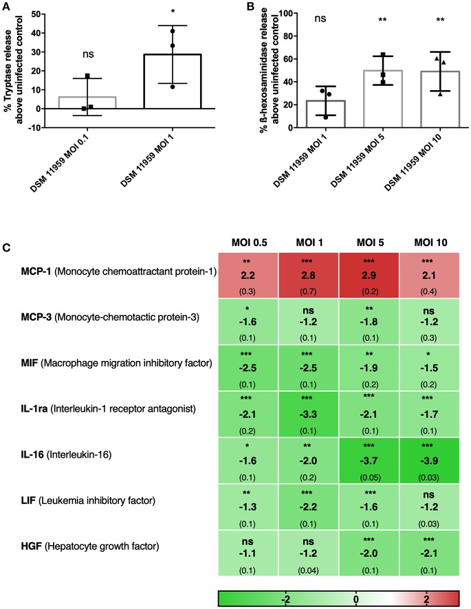
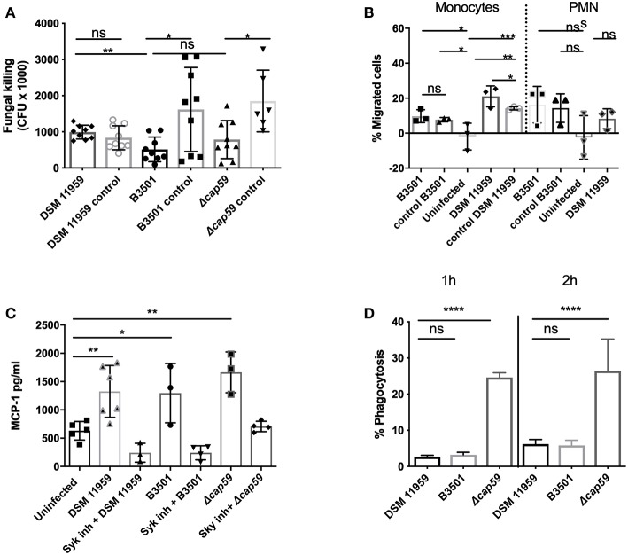
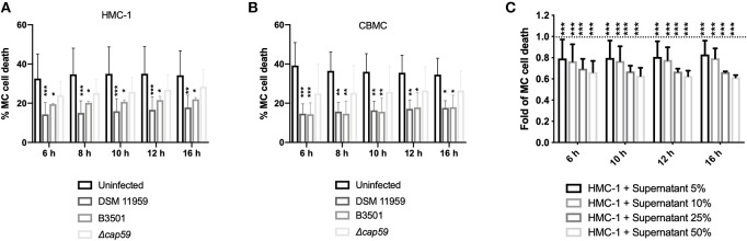
Cryptococcosis, caused by the basidiomycete Cryptococcus neoformans, is a life-threatening disease affecting approximately one million people per year worldwide. Infection can occur when C. neoformans cells are inhaled by immunocompromised people. In order to establish infection, the yeast must bypass recognition and clearance by immune cells guarding the tissue. Using in vitro infections, we characterized the role of mast cells (MCs) in cryptococcosis. We found that MCs recognize C. neoformans and release inflammatory mediators such as tryptase and cytokines. From the latter group MCs released mainly CCL-2/MCP-1, a strong chemoattractant for monocytic cells. We demonstrated that supernatants of infected MCs recruit monocytes but not neutrophils. During infection with C. neoformans, MCs have a limited ability to kill the yeast depending on the serotype. C. neoformans, in turn, modulates the lifespan of MCs both, by presence of its polysaccharide capsule and by secreting soluble modulators. Taken together, MCs might have important contributions to fungal clearance during early stages of cryptocococis where these cells regulate recruitment of monocytes to mucosal tissues.

Keywords: Cryptococcus neoformans; fungi; innate immunity; mast cells; monocyte chemoattractant protein 1/CCL-2; monocytes.


Figures

Figure 1
Figure 1
MCs show direct action against C. neoformans and release granular proteins. (A) MCs released tryptase after 6 h of infection with C. neoformans (MOI 1). Data from n = 3 (3). (B) MCs released ß-hexosaminidase after 1 h infection with C. neoformans (MOI 5 and 10). Data from n = 3 (3). (C) Fold change of a total of 48 cytokines were measured in supernatants of MCs infected with C. neoformans at different MOIs. Seven cytokines were differentially expressed and only MCP-1 was upregulated. Fold change was calculated as the ratio between infected condition and uninfected control. Data from n = 2 (4). For all analyses significance was analyzed by multi-comparison using one-way ANOVA with Bonferroni post-test. *P ≤ 0.05; **P ≤ 0.01; ***P ≤ 0.001; ns, not significant.
Figure 2
Figure 2
MC recognition of C. neoformans was mediated by Syk signaling and chemoattracts monocytes. (A) MCs induced killing was serotype dependent. Serotype A (DSM 11959) resisted killing by MCs whereas serotype D (B3501) and acapsular mutant cap59 showed some level of susceptibility. Data from n = 9. (B) Monocytes but not neutrophils were recruited by MCs after C. neoformans infection. The graph depicts the percentage of migration of neutrophil or monocyte migration over time toward supernatants of MCs infected with C. neoformans. (C) MCs secreted MCP-1 in a Syk dependent manner after 20 h infection with C. neoformans (MOI 5). MCP-1 was measured by ELISA in supernatants of HMC-1 infected with C. neoformans (MOI 5). Data from n = 3 (3). Syk was inhibited by piceatannol 10 μM after infection with HMC-1 infection with C. neoformans strains. (D) Acapsular mutants induce significantly more phagocytosis after 1 and 2 h infection. MC phagocytosis of C. neoformans was determined by FACS as the percentage of IgG-Alexa-488 dye high/IgG-Alexa-633 low Cryptococcus cells. Data from n = 4. Significance was analyzed by multi-comparison using one-way ANOVA with Bonferroni post-test in (B,D). For (A,C) significance was analyzed using unpaired t-test with Welch correction. *P ≤ 0.05; **P ≤ 0.01; ***P ≤ 0.001; ****P ≤ 0.0001; ns, not significant.
Figure 3
Figure 3
C. neoformans infection increase MC survival. (A) Infection with capsular C. neoformans increased HMC-1 survival. Infections were performed using MOI of 5. Data from n = 6 (3). (B) Infection with capsular C. neoformans (MOI 5) increased viability of primary MCs. Data from n = 4 (3). (C) Secreted components from C. neoformans overnight cultures induced MC survival. Plotted is the delayed death of resting MCs incubated with increasing concentrations of supernatants of an overnight culture of C. neoformans plotted as fold change in relation to untreated MCs (dashed line). For all analyses significance was analyzed by multi-comparison using two-way ANOVA with Bonferroni post-test. *P ≤ 0.05; **P ≤ 0.01; ***P ≤ 0.001; ****P ≤ 0.0001; ns, not significant.

Similar articles

Cited by

References

    1. Agier J., Pastwinska J., Brzezinska-Błaszczyk E. (2018). An overview of mast cell pattern recognition receptors. Inflamm. Res. 67, 737–746. 10.1007/s00011-018-1164-5 - DOI - PMC - PubMed
    1. Alanio A., Desnos-Ollivier M., Dromer F. (2011). Dynamics of Cryptococcus neoformans-macrophage interactions reveal that fungal background influences outcome during cryptococcal meningoencephalitis in humans. MBio 2:e00158–11. 10.1128/mBio.00158-11 - DOI - PMC - PubMed
    1. Alspaugh J. A. (2015). Virulence mechanisms and Cryptococcus neoformans pathogenesis. Fungal Genet. Biol. 78, 55–58. 10.1016/j.fgb.2014.09.004 - DOI - PMC - PubMed
    1. Barbosa-Lorenzi V. C., Peyda S., Scheynius A., Nilsson G., Lunderius-Andersson C. (2017). Curdlan induces selective mast cell degranulation without concomitant release of LTC4, IL-6 or CCL2. Immunobiology 222, 647–650. 10.1016/j.imbio.2016.12.001 - DOI - PubMed
    1. Beghdadi W., Madjene L. C., Benhamou M., Charles N., Gautier G., Launay P., et al. . (2011). Mast cells as cellular sensors in inflammation and immunity. Front. Immunol. 2:37. 10.3389/fimmu.2011.00037 - DOI - PMC - PubMed

Publication types

MeSH terms