

Effect of a Controlled Ankle Motion Walking Boot on Syndesmotic Instability During Weightbearing: A Cadaveric Study
- PMID: 31457067
- PMCID: PMC6702776
- DOI: 10.1177/2325967119864018
Effect of a Controlled Ankle Motion Walking Boot on Syndesmotic Instability During Weightbearing: A Cadaveric Study
Abstract
Background: Syndesmotic injuries can lead to long-term complications; hence, they require careful management. Conservative treatment is adequate when 1 syndesmotic ligament is injured, but surgery is often necessary to achieve articular congruity when 3 syndesmotic ligaments are ruptured. However, there is some controversy over the best treatment for 2-ligament injuries.
Purpose: To evaluate the effect of a controlled ankle motion (CAM) walking boot on syndesmotic instability following iatrogenic isolated anterior inferior tibiofibular ligament (AiTFL) injury and combined AiTFL/interosseous ligament (IOL) injuries in a cadaveric simulated weightbearing model.
Study design: Controlled laboratory study.
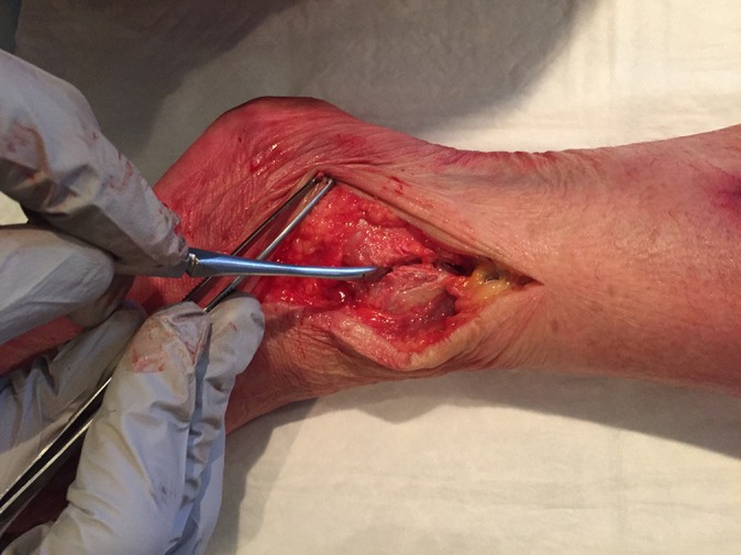
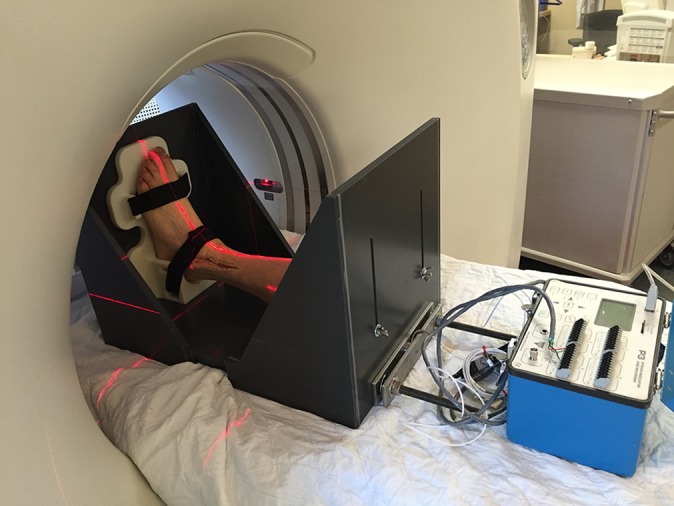
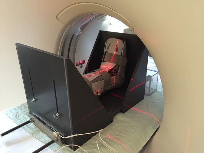
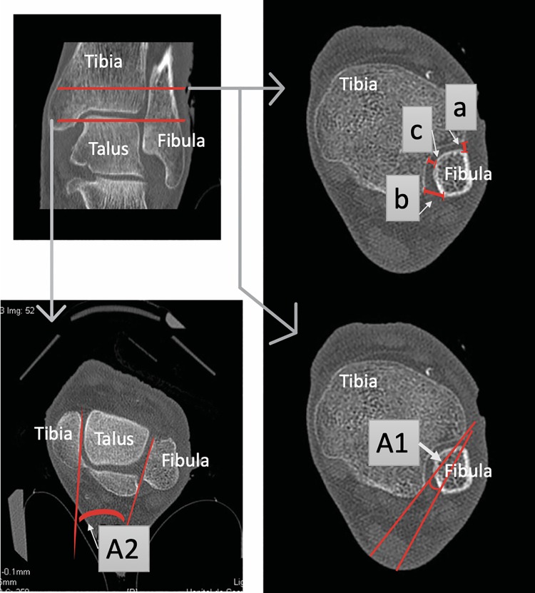
Methods: Ten cadaveric specimens were dissected to expose the tibial plateau and syndesmosis. The specimens were fitted to a custom-made device, and a reproducible axial load of 750 N was applied. Iatrogenic rupture of the syndesmotic ligaments (AiTFL + IOL) was done sequentially. Uninjured syndesmoses, isolated AiTFL rupture, and combined AiTFL/IOL rupture were compared with and without axial loading (AL) and CAM boot. The distal tibiofibular relationship was evaluated using a previously validated computed tomography scan measurement system. Wilcoxon tests for paired samples and nonparametric data were used.
Results: The only difference noted in the distal tibiofibular relationship during AL was an increase in the external rotation of the fibula when using the CAM boot. This was observed with AiTFL rupture (8.40° vs 11.17°; P = .009) and combined AiTFL/IOL rupture (8.81° vs 11.97°; P = .005).
Conclusion: AL did not cause a significant displacement between the tibia and fibula, even when 2 ligaments were ruptured. However, the CAM boot produced a significant external rotation with 1 or 2 injured ligaments.
Clinical relevance: Further studies are needed to assess the capacity of the CAM walking boot to prevent malreduction when external rotation forces are applied to the ankle. Moreover, special care should be taken during the fitting of the CAM boot to avoid overinflation of the cushions.
Keywords: CAM orthopaedic boot; high ankle sprain; syndesmotic injury; weightbearing.
Conflict of interest statement
One or more of the authors has declared the following potential conflict of interest or source of funding: This study was supported by an educational grant by the PREMIER program. J.H.-D. has received consulting fees from Globus Medical and DePuy Synthes. S.L. has received consulting fees from Stryker. É.S. has a family member who is employed by Smith & Nephew. The Hôpital du Sacré-Cœur de Montréal has received educational and research support from Arthrex, ConMed, DePuy, Linvatec, Medacta, Smith & Nephew, Stryker, Synthes, Tornier, Wright, and Zimmer Biomet. AOSSM checks author disclosures against the Open Payments Database (OPD). AOSSM has not conducted an independent investigation on the OPD and disclaims any liability or responsibility relating thereto.
Figures
References
-
- Amendola A, Williams G, Foster D. Evidence-based approach to treatment of acute traumatic syndesmosis (high ankle) sprains. Sports Med Arthrosc Rev. 2006;14(4):232–236. - PubMed
-
- Beumer A, Valstar ER, Garling EH. et al. Effects of ligament sectioning on the kinematics of the distal tibiofibular syndesmosis: a radiostereometric study of 10 cadaveric specimens based on presumed trauma mechanisms with suggestions for treatment. Acta Orthop. 2006;77(3):531–540. - PubMed
-
- Beumer A, Campo MM, Niesing R, Day J, Kleinrensink G-J, Swierstra BA. Screw fixation of the syndesmosis: a cadaver model comparing stainless steel and titanium screws and three and four cortical fixation. Injury. 2005;36(1):60–64. - PubMed
-
- Boytim MJ, Fischer DA, Neumann L. Syndesmotic ankle sprains. Am J Sports Med. 1991;19(3):294–298. - PubMed
-
- Close JR. Some applications of the functional anatomy of the ankle joint. J Bone Joint Surg Am. 1956;38(4):761–781. - PubMed
LinkOut - more resources
Full Text Sources
