. 2019 Jun 24;4(6):10906-10914.
doi: 10.1021/acsomega.9b00756. eCollection 2019 Jun 30.

Crystal Structure of FOXC2 in Complex with DNA Target

Affiliations

Crystal Structure of FOXC2 in Complex with DNA Target

Shichang Li et al. ACS Omega. .

Abstract

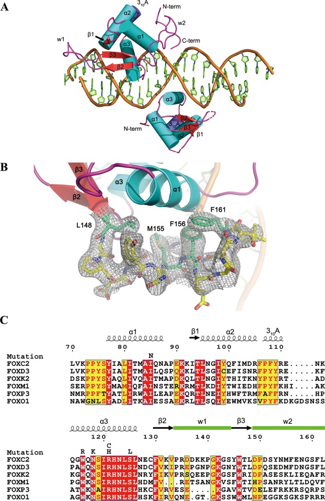
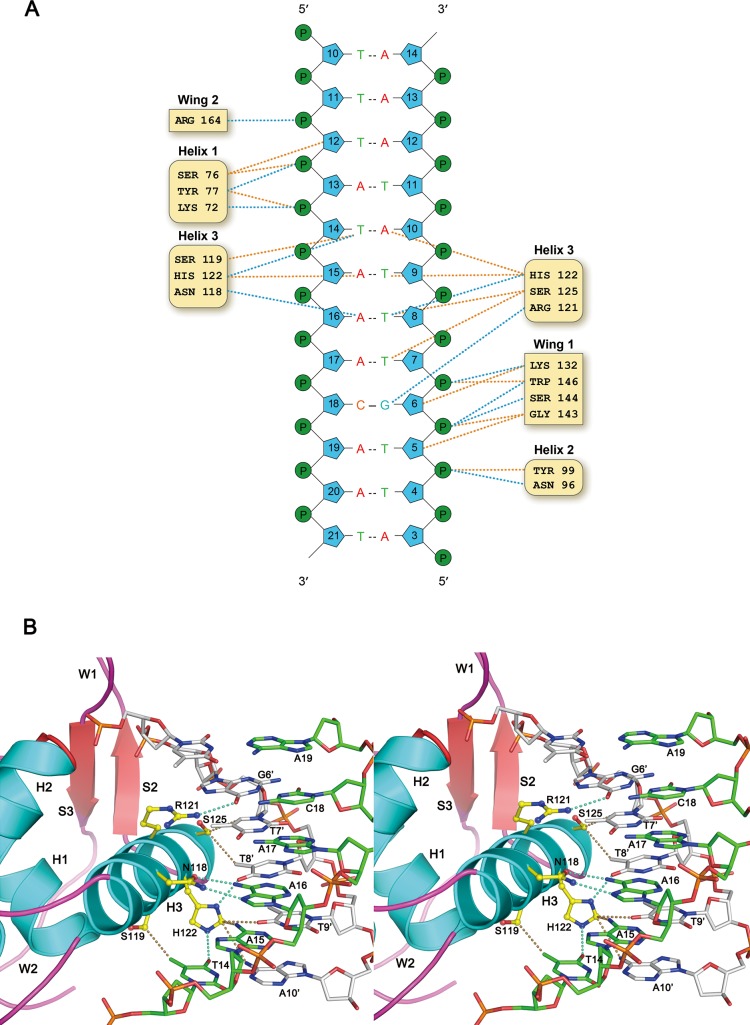
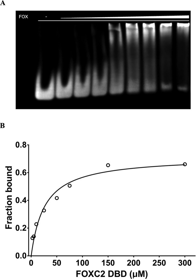
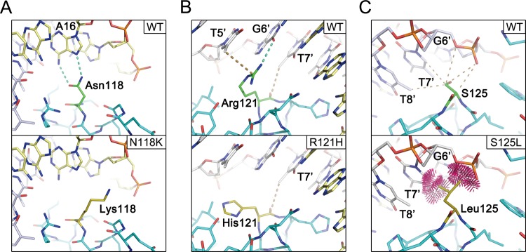
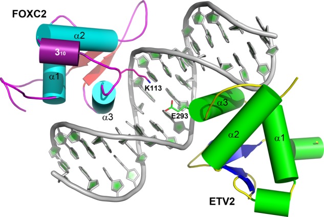
Forkhead transcription factor C2 (FOXC2) is a transcription factor regulating vascular and lymphatic development, and its mutations are linked to lymphedema-distichiasis syndrome. FOXC2 is also a crucial regulator of the epithelial-mesenchymal transition processes essential for tumor metastasis. Here, we report the crystal structure of the FOXC2-DNA-binding domain in complex with its cognate DNA. The crystal structure provides the basis of DNA sequence recognition by FOXC2 for the T/CAAAC motif. Helix 3 makes the majority of the DNA-protein interactions and confers the DNA sequence specificity. The computational energy calculation results also validate the structural observations. The FOXC2 and DNA complex structure provides a detailed picture of protein and DNA interactions, which allows us to predict its DNA recognition specificity and impaired functions in mutants identified in human patients.


Conflict of interest statement

The authors declare no competing financial interest.

Figures

Figure 1
Figure 1
Overall structure of the FOXC2 DBD–DNA complex and sequence alignment. (A) A schematic of the protein–DNA complex. The FOXC2 DBD is indicated in cyan (helices), red (sheets), and magenta (coils). The DNA containing dual binding sites of FOXC2 are also illustrated in cartoon representation. The secondary structure elements and N- and C-termini are labeled. The dotted lines represent the loops missing in the Mol B model. (B) C-terminal residues of FOXC2, 148–161, are depicted in the stick representation with a 2FoFc map (1σ level). (C) Sequence alignment of the FOX family DNA-binding domains. The numbering is based on the FOXC2 residues. The secondary structure elements are indicated, and the missense mutations identified in FOXC2 are also denoted on top of the sequences.
Figure 2
Figure 2
FOXC2 DBD and DNA interaction. (A) NuProPlot diagram of FOXC2 DBD–DNA interactions. The DNA segment interacting with the Mol A is illustrated. Hydrogen bonds and van der Waals interactions are represented by dotted blue and orange lines, respectively. (B) Detailed view of the FOXC2 DBD and its cognate DNA segment shown in a stereo diagram. The side chains of amino acid residues involved in the DNA base interactions are illustrated with the carbons in yellow, the oxygens in red, and the nitrogens in blue.
Figure 3
Figure 3
Electrophoretic mobility shift assay of FOXC2 and DNA. (A) FOXC2 and DNA binding by EMSA. The purified FOXC2 DBD was mixed with the DNA containing TAAACA motif, and the protein–DNA complexes were separated on a 6% acrylamide gel. Increasing concentrations of FOXC2 DBD, 0, 2.5, 5, 10, 25, 50, 75, 150, and 300 μM, were used (lanes 1–9). (B) Linear scale saturation binding curve of FOXC2 DBD measured by EMSA. The Kd and R2 were estimated as 26.4 ± 3.9 μM (95% CI: 18.7–37 μM) and 0.94, respectively. The error bar indicates a standard deviation of measurements from triplicate experiments.
Figure 4
Figure 4
Comparisons of wild type and FOXC2 missense mutants. Models of wild type, N118K (A), R121H (B), and S125L (C), are depicted in the stick representation. Dotted lines represent hydrogen bonds, and in panel (C), the clash among the atoms is illustrated.
Figure 5
Figure 5
Composite model of FOXC2 and ETV2 bound to a DNA target. A ternary complex of FOXC2 and ETV2 bound to a FOX–ETS motif is illustrated. The FOXC2 follows the same color scheme as in Figure 1A, and ETV2 is indicated in green (helices), blue (sheets), and yellow (coils). The FOX–ETS DNA is illustrated in the cartoon representation. Two charged residues at the interface, Lys113 of FOXC2 and Glu293 of ETV2, are labeled.

References

    1. Mani S. A.; Guo W.; Liao M.-J.; Eaton E. N.; Ayyanan A.; Zhou A. Y.; Brooks M.; Reinhard F.; Zhang C. C.; Shipitsin M.; Campbell L. L.; Polyak K.; Brisken C.; Yang J.; Weinberg R. A. The epithelial-mesenchymal transition generates cells with properties of stem cells. Cell 2008, 133, 704–715. 10.1016/j.cell.2008.03.027. - DOI - PMC - PubMed
    1. Mani S. A.; Yang J.; Brooks M.; Schwaninger G.; Zhou A.; Miura N.; Kutok J. L.; Hartwell K.; Richardson A. L.; Weinberg R. A. Mesenchyme Forkhead 1 (FOXC2) plays a key role in metastasis and is associated with aggressive basal-like breast cancers. Proc. Natl. Acad. Sci. U.S.A. 2007, 104, 10069–10074. 10.1073/pnas.0703900104. - DOI - PMC - PubMed
    1. Hollier B. G.; Tinnirello A. A.; Werden S. J.; Evans K. W.; Taube J. H.; Sarkar T. R.; Sphyris N.; Shariati M.; Kumar S. V.; Battula V. L.; Herschkowitz J. I.; Guerra R.; Chang J. T.; Miura N.; Rosen J. M.; Mani S. A. FOXC2 expression links epithelial-mesenchymal transition and stem cell properties in breast cancer. Cancer Res. 2013, 73, 1981–1992. 10.1158/0008-5472.CAN-12-2962. - DOI - PMC - PubMed
    1. De Craene B.; Berx G. Regulatory networks defining EMT during cancer initiation and progression. Nat. Rev. Cancer 2013, 13, 97–110. 10.1038/nrc3447. - DOI - PubMed
    1. Kume T. The Role of FoxC2 Transcription Factor in Tumor Angiogenesis. J. Oncol. 2012, 2012, 20459310.1155/2012/204593. - DOI - PMC - PubMed