Review
. 2019 Sep 17;52(9):2582-2597.
doi: 10.1021/acs.accounts.9b00302. Epub 2019 Aug 28.

Reaction-Based Fluorescent Probes for the Detection and Imaging of Reactive Oxygen, Nitrogen, and Sulfur Species

Affiliations
Review

Reaction-Based Fluorescent Probes for the Detection and Imaging of Reactive Oxygen, Nitrogen, and Sulfur Species

Luling Wu et al. Acc Chem Res. .

Abstract

This Account describes a range of strategies for the development of fluorescent probes for detecting reactive oxygen species (ROS), reactive nitrogen species (RNS), and reactive (redox-active) sulfur species (RSS). Many ROS/RNS have been implicated in pathological processes such as Alzheimer's disease, cancer, diabetes mellitus, cardiovascular disease, and aging, while many RSS play important roles in maintaining redox homeostasis, serving as antioxidants and acting as free radical scavengers. Fluorescence-based systems have emerged as one of the best ways to monitor the concentrations and locations of these often very short lived species. Because of the high levels of sensitivity and in particular their ability to be used for temporal and spatial sampling for in vivo imaging applications. As a direct result, there has been a huge surge in the development of fluorescent probes for sensitive and selective detection of ROS, RNS, and RSS within cellular environments. However, cellular environments are extremely complex, often with more than one species involved in a given biochemical process. As a result, there has been a rise in the development of dual-responsive fluorescent probes (AND-logic probes) that can monitor the presence of more than one species in a biological environment. Our aim with this Account is to introduce the fluorescent probes that we have developed for in vitro and in vivo measurement of ROS, RNS, and RSS. Fluorescence-based sensing mechanisms used in the construction of the probes include photoinduced electron transfer, intramolecular charge transfer, excited-state intramolecular proton transfer (ESIPT), and fluorescence resonance energy transfer. In particular, probes for hydrogen peroxide, hypochlorous acid, superoxide, peroxynitrite, glutathione, cysteine, homocysteine, and hydrogen sulfide are discussed. In addition, we describe the development of AND-logic-based systems capable of detecting two species, such as peroxynitrite and glutathione. One of the most interesting advances contained in this Account is our extension of indicator displacement assays (IDAs) to reaction-based indicator displacement assays (RIAs). In an IDA system, an indicator is allowed to bind reversibly to a receptor. Then a competitive analyte is introduced into the system, resulting in displacement of the indicator from the host, which in turn modulates the optical signal. With an RIA-based system, the indicator is cleaved from a preformed receptor-indicator complex rather than being displaced by the analyte. Nevertheless, without a doubt the most significant result contained in this Account is the use of an ESIPT-based probe for the simultaneous sensing of fibrous proteins/peptides AND environmental ROS/RNS.


Conflict of interest statement

The authors declare no competing financial interest.

Figures

Scheme 1
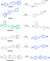
Scheme 1. (a) 1 and 1d-Fructose Complex for Detecting H2O2; (b) 2 and 2d-Fructose Complex for Detecting H2O2
Scheme 2
Scheme 2. Boronate Probes for Detecting H2O2
Scheme 3
Scheme 3. Mechanism and Fluorescence Spectra of the ARS–PBA Complex for Detecting H2O2
Adapted from ref (35). Published by The Royal Society of Chemistry.
Scheme 4
Scheme 4. ESIPT-Based Probe TCBT-OMe for Detecting HOCl/ClO
Scheme 5
Scheme 5. ESIPT-Based Probe C7 for Detecting HOCl
Scheme 6
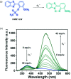
Scheme 6. (a) ESIPT-Based Probe HMBT-LW for Detecting O2•–; (b) Changes in Fluorescence Emission Intensity of HMBT-LW (5 μM) with Increasing O2•– in Phosphate-Buffered Saline (PBS) (10 mM v/v, 1:1 DMSO/PBS, pH 7.4) after 3 min at λex = 310 nm
Adapted with permission from ref (40). Published by The Royal Society of Chemistry (RSC) on behalf of the Centre National de la Recherche Scientifique (CNRS) and the RSC.
Scheme 7
Scheme 7. Probe 3 for Detecting ONOO in the Presence of d-Fructose
Scheme 8
Scheme 8. ARS–NBA Complex for Detecting ONOO
Figure 1
Figure 1
Probe 4 for detecting ONOO.
Scheme 9
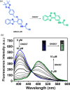
Scheme 9. (a) Probe ABAH-LW for Detecting ONOO; (b) Changes in Fluorescence Emission of ABAH-LW (3 μM) with Increasing Addition of ONOO in PBS (pH 8.2, Containing 8% DMSO, 1 mM CTAB) after 1 min at λex = 370 nm
Adapted with permission from ref (46). Copyright 2018 Royal Society of Chemistry.
Figure 2
Figure 2
TCFB1 and TCFB2 for detecting ONOO.
Scheme 10
Scheme 10. PR1 for Detecting ONOO
Scheme 11
Scheme 11. 1–copper(II) Complex for Detecting NO and HNO
Scheme 12
Scheme 12. Probe 5 for Detecting GSH
Figure 3
Figure 3
TCF-GSH and TCFCl-GSH for detecting GSH.
Figure 4
Figure 4
Theranostic prodrug DCM-S-CPT.
Figure 5
Figure 5
(a) Probe 6 for detecting thiols. (b) Fluorescence emission spectra of probe 6 (10 μM) before and after addition of l-cysteine (300 μM) in 4:1 v/v MeOH/H2O at λex = 505 nm at 37 °C. Adapted with permission from ref (55). Copyright 2012 Royal Society of Chemistry.
Scheme 13
Scheme 13. DT-Gal for Detecting H2S
Scheme 14
Scheme 14. OPD for Detecting Na2S
Scheme 15
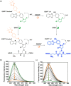
Scheme 15. (a) GSH-PF3 for Detecting ONOO AND GSH; (b, c) Fluorescence Spectra of GSH-PF3 (0.5 μM) upon Addition of (a) ONOO (10 μM) Followed by GSH (0–80 μM) with a 5 min Wait between Additions and (c) GSH (200 μM) Followed by Addition of ONOO (0–10 μM) with a 10 min Wait between Additions (52 wt % Methanol, pH 8.21, λex = 488 nm, 25 °C)
Adapted with permission from ref (59). Copyright 2018 Royal Society of Chemistry.
Scheme 16
Scheme 16. (a) GSH-ABAH for Detecting ONOO AND GSH; (b, c) Fluorescence Spectra of GSH-ABAH (2 μM) upon Addition of (a) ONOO (4 μM) Followed by GSH (0–2 μM) after a 1 min Wait and (c) GSH (5 μM) Followed by ONOO (0–14 μM) after a 1 min Wait (8% DMSO, 1 mM CTAB, pH 8.20, λex = 390 nm, 25 °C)
Adapted with permission from ref (60). Copyright 2018 Royal Society of Chemistry.
Scheme 17
Scheme 17. Pinkment-OH for Detecting ONOO, Pinkment-OTBS for Detecting ONOO AND Fluoride, and Pinkment-OAc for Detecting ONOO AND Esterase Activity
Scheme 18
Scheme 18. Dual-Enzyme-Activated PF3-Glc
Figure 6
Figure 6
(a) ESIPT probes 3-HF-X (X = OMe, Me, H) for detecting ONOO. The normal (N) and phototautomeric (T*) forms are shown. (b, c) Fluorescence imaging of a brain section of a transgenic mouse treated with 3-HF-OMe (20 μM) (b) without and (c) with ONOO (30 μM). The excitation/emission wavelengths for the blue (N-state), green (T*-state), and red (anti-Aβ antibody) channels are 404/425–475, 404/500–550, and 561/640–730 nm, respectively. The white arrows indicate stained Aβ aggregates. Reprinted from ref (64). Copyright 2018 American Chemical Society.

References

    1. Valko M.; Leibfritz D.; Moncol J.; Cronin M. T. D.; Mazur M.; Telser J. Free radicals and antioxidants in normal physiological functions and human disease. Int. J. Biochem. Cell Biol. 2007, 39, 44–84. 10.1016/j.biocel.2006.07.001. - DOI - PubMed
    1. Winyard P. G.; Ryan B.; Eggleton P.; Nissim A.; Taylor E.; Lo Faro M. L.; Burkholz T.; Szabó-Taylor K. E.; Fox B.; Viner N.; Haigh R. C.; Benjamin N.; Jones A. M.; Whiteman M. Measurement and meaning of markers of reactive species of oxygen, nitrogen and sulfur in healthy human subjects and patients with inflammatory joint disease. Biochem. Soc. Trans. 2011, 39 (5), 1226–1232. 10.1042/BST0391226. - DOI - PubMed
    1. Yin C.-X.; Xiong K.-M.; Huo F.-J.; Salamanca J. C.; Strongin R. M. Fluorescent Probes with Multiple Binding Sites for the Discrimination of Cys, Hcy, and GSH. Angew. Chem., Int. Ed. 2017, 56, 13188–13198. 10.1002/anie.201704084. - DOI - PMC - PubMed
    1. Mishanina T. V.; Libiad M.; Banerjee R. Biogenesis of reactive sulfur species for signaling by hydrogen sulfide oxidation pathways. Nat. Chem. Biol. 2015, 11, 457.10.1038/nchembio.1834. - DOI - PMC - PubMed
    1. Stamler J. S.; Slivka A. Biological chemistry of thiols in the vasculature and in vascular-related disease. Nutr. Rev. 1996, 54, 1–30. 10.1111/j.1753-4887.1996.tb03770.x. - DOI - PubMed

Publication types

MeSH terms