Practical Guidance for Measuring and Reporting Surgical Margins in Vulvar Cancer
- PMID: 31460873
- PMCID: PMC7478209
- DOI: 10.1097/PGP.0000000000000631
Practical Guidance for Measuring and Reporting Surgical Margins in Vulvar Cancer
Abstract
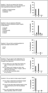
Surgical resection with free surgical margins is the cornerstone of successful primary treatment of vulvar squamous cell carcinoma (VSCC). In general reexcision is recommended when the minimum peripheral surgical margin (MPSM) is <8 mm microscopically. Pathologists are, therefore, required to report the minimum distance from the tumor to the surgical margin. Currently, there are no guidelines on how to make this measurement, as this is often considered straightforward. However, during the 2018 Annual Meeting of the British Association of Gynaecological Pathologists (BAGP), a discussion on this topic revealed a variety of opinions with regard to reporting and method of measuring margin clearance in VSCC specimens. Given the need for uniformity and the lack of guidance in the literature, we initiated an online survey in order to deliver a consensus-based definition of peripheral surgical margins in VSCC resections. The survey included questions and representative diagrams of peripheral margin measurements. In total, 57 pathologists participated in this survey. On the basis of consensus results, we propose to define MPSM in VSCC as the minimum distance from the peripheral edge of the invasive tumor nests toward the inked peripheral surgical margin reported in millimeters. This MPSM measurement should run through tissue and preferably be measured in a straight line. Along with MPSM, other relevant measurements such as depth of invasion or tumor thickness and distance to deep margins should be reported. This manuscript provides guidance to the practicing pathologist in measuring MPSM in VSCC resection specimens, in order to promote uniformity in measuring and reporting.
Conflict of interest statement
K.J.P. is funded by the NIH/NCI Institutional Support Grant P30 CA008748. The authors declare no conflict of interest.
Figures


References
-
- Hacker NF, Eifel PJ, van der Velden J. Cancer of the vulva. Int J Gynaecol Obstet 2012;119(suppl 2):S90–6. - PubMed
-
- Te Grootenhuis NC, van der Zee AG, van Doorn HC, et al. Sentinel nodes in vulvar cancer: long-term follow-up of the GROningen INternational Study on Sentinel nodes in Vulvar cancer (GROINSS-V) I. Gynecol Oncol 2016;140:8–14. - PubMed
-
- Luesley DM, Barton DPJ, Bishop M, et al. Guidelines for the diagnosis and management of vulval carcinoma. 2014. Available at: www.rcog.org.uk/globalassets/documents/guidelines/vulvalcancerguideline.pdf. Accessed April 17, 2019.
-
- National Dutch guideline gynaecologic tumors; vulvar carcinoma. 2015. Available at: www.oncoline.nl/vulvacarcinoom. Accessed April 17, 2019.
-
- Heaps JM, Fu YS, Montz FJ, et al. Surgical-pathologic variables predictive of local recurrence in squamous cell carcinoma of the vulva. Gynecol Oncol 1990;38:309–14. - PubMed
MeSH terms
Grants and funding
LinkOut - more resources
Full Text Sources
Medical

