. 2019 Nov 1;317(5):F1201-F1210.
doi: 10.1152/ajprenal.00629.2018. Epub 2019 Aug 28.

Inactivation of Tsc2 in Abcg2 lineage-derived cells drives the appearance of polycystic lesions and fibrosis in the adult kidney

Affiliations

Inactivation of Tsc2 in Abcg2 lineage-derived cells drives the appearance of polycystic lesions and fibrosis in the adult kidney

Leslie S Gewin et al. Am J Physiol Renal Physiol. .

Abstract

Tuberous sclerosis complex 2 (TSC2), or tuberin, is a pivotal regulator of the mechanistic target of rapamycin signaling pathway that controls cell survival, proliferation, growth, and migration. Loss of Tsc2 function manifests in organ-specific consequences, the mechanisms of which remain incompletely understood. Recent single cell analysis of the kidney has identified ATP-binding cassette G2 (Abcg2) expression in renal proximal tubules of adult mice as well as a in a novel cell population. The impact in adult kidney of Tsc2 knockdown in the Abcg2-expressing lineage has not been evaluated. We engineered an inducible system in which expression of truncated Tsc2, lacking exons 36-37 with an intact 3' region and polycystin 1, is driven by Abcg2. Here, we demonstrate that selective expression of Tsc2fl36-37 in the Abcg2pos lineage drives recombination in proximal tubule epithelial and rare perivascular mesenchymal cells, which results in progressive proximal tubule injury, impaired kidney function, formation of cystic lesions, and fibrosis in adult mice. These data illustrate the critical importance of Tsc2 function in the Abcg2-expressing proximal tubule epithelium and mesenchyme during the development of cystic lesions and remodeling of kidney parenchyma.

Keywords: ATP-binding cassette G2; polycystic kidney disease; tuberous sclerosis complex 2.


Conflict of interest statement

No conflicts of interest, financial or otherwise, are declared by the authors.

Figures
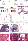
Fig. 1.
Fig. 1.
Progressive cyst formation and fibrosis in the adult kidney after expression of truncated tuberous sclerosis complex 2 (Tsc2). A: study timeline. Adult female and male mice were induced between 8 and 10 wk of age with low-dose tamoxifen and euthanized at 12 or 20 wk. Groups were as follows: ATP-binding cassette G2 (Abcg2)CreERT2 Tsc2fl36–37+/+, +/−, or −/−. T, time; HCT, hematocrit. B and C: representative gross anatomy of kidneys correlated to genotype and functional end points (20 wk). WT, wild type. D: hemotyostain of cystic kidney tissue. Scale bar = 100 μm. E: mean body weight. F: mean kidney weight. G: mean kidney length at 20 wk. n = 5+/+ and 6−/− female mice and n = 6+/+ and 6−/− male mice. H, I, K, and M: trichrome stain of Tsc2+/+ and Tsc2−/− kidney tissue (20 wk). Collagen is visualized as blue. Scale bars = 50 μm. J: collagen type I-α1 (Col1A1) expression was quantitated by PCR using total RNA isolated from female and male kidney tissue from 12 and 20 wk. n = 6+/+ and 8−/− female mice and n = 6+/+ and 7−/− male mice. Data are presented as means ± SE. Significance was determined using one-way ANOVA with a Tukey’s post hoc test.
Fig. 2.
Fig. 2.
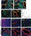
Loss of tuberous sclerosis complex 2 (Tsc2) function in the adult ATP-binding cassette G2-positive (Abcg2pos) kidney lineage drives mechanistic target of rapamycin (mTOR) activation, proximal tubule injury, and myofibroblast accumulation. Adult female and male mice were induced between 8 and 10 wk of age with tamoxifen and euthanized at 12 or 20 wk. Histological evaluation and lineage analysis were performed using Tsc2+/+ and Tsc2−/− kidneys. A: tetragonolobus lectin (LTL) colocalization of the proximal tubule epithelium with enhanced green fluorescent protein (eGFP). B: enlarged area from A. Scale bars = 50 μm. C and D: staining to localize eGFP (green) and pS6 (red) expression as an indication of mTOR signaling. Nuclei were visualized with DAPI. Scale bars = 100 μm. E: kidney injury molecule-1 (Kim1) expression was quantitated by PCR using total RNA isolated from female and male kidney tissue from 12 and 20 wk. n = 6+/+ and 8−/− female mice and n = 6+/+ and 7−/− male mice. Data are presented as means ± SE. Significance was determined using one-way ANOVA with a Tukey’s post hoc. F−K: immunofluorescent staining to localize α-smooth muscle actin (α-SMA; green) and eGFP (red) expression. G and H: eGFP localization in the cortex or medulla. WT, wild type. Magnification: ×4 (scale bar = 100 μm) and ×40 (scale bar = 50 μm).
Fig. 3.
Fig. 3.
Impaired function is evident after expression of truncated tuberous sclerosis complex 2 (Tsc2 ) in ATP-binding cassette G2 (Abgc2) linage cells. A: blood urea nitrogen (BUN) was measured in mouse serum. n = 7+/+ and 9−/− female mice and n = 15+/+ and 7−/− male mice. B: the albumin-to-creatinine ratio was measured in urine. n = 13+/+ and 12−/− female mice and n = 11+/+ and 7−/− male mice. C: hematocrit was measured using a standard protocol. n = 8+/+ and 10−/− female mice and n = 8+/+ and 6−/− male mice. Groups were pooled from 12- and 20-wk time points. D: mean systemic blood pressure at 20 wk. n = 11+/+ and 8−/− mice. E: Fulton’s index (right ventricle/left ventricle + septum). n = 16+/+ and 26−/− mice. WT, wild type. Data are presented as means ± SE. Significance was determined using one-way ANOVA with a Tukey’s post hoc test.
Fig. 4.
Fig. 4.
Knockdown of tuberous sclerosis complex 2 (Tsc2) induces epithelial injury that precedes fibrosis. A: adult female and male mice were induced between 8 and 10 wk of age with tamoxifen and euthanized at 3, 6, and 9 wk. Groups were as follows: ATP-binding cassette G2 (Abcg2)CreERT2 Tsc2fl36–37+/+ or −/−. T, time; HCT, hematocrit. BG: representative hematoxylin and eosin staining of kidney tissue. Scale bars = 100 μm. H: HCT was measured using a standard protocol. n = 8+/+ and 3−/− female mice and n = 4+/+ and 2−/− male mice at 3 wk. I: mean kidney weight. J: mean kidney length. 3 wk: n = 6 female mice (3+/+ and 3−/−) and 6 male mice (3+/+ and 3−/−); 6 wk: n = 6 female mice (3+/+ and 3−/−) and 7 male mice (3+/+ and 4−/−); 9 wk: n = 6 female mice (3+/+ and 3−/−) and 7 male mice (3+/+ and 4−/−). Data are presented as means ± SE. Significance was determined using one-way ANOVA with a Tukey’s post hoc test.
Fig. 5.
Fig. 5.
Progressive fibrosis was associated with knockdown of tuberous sclerosis complex 2 (Tsc2) in the ATP-binding cassette G2 (ABCG2) lineage. AD: representative trichrome staining of Tsc2fl36–37+/+ or −/− adult kidney tissue. Scale bars = 100 μm. Arrows indicate interstitial fibrosis.
Fig. 6.
Fig. 6.
Progressive cyst formation driven by mesodermal tuberous sclerosis complex 2 (Tsc2) depletion during development is accompanied by interstitial fibrosis. With the use of the Dermo1Cre driver, Tsc2 was depleted in the mesoderm and derivatives during development. Mice were euthanized on postnatal days 7, 18, and 21 (P7, P18, and P21, respectively). AC: representative trichrome-stained kidneys. DG: enlarged areas of P18 and P21 indicated by boxes. Arrows indicate interstitial fibrosis. WT, wild type.

References

    1. Asakura A, Rudnicki MA. Side population cells from diverse adult tissues are capable of in vitro hematopoietic differentiation. Exp Hematol 30: 1339–1345, 2002. doi:10.1016/S0301-472X(02)00954-2. - DOI - PubMed
    1. Bissler JJ, Zadjali F, Bridges D, Astrinidis A, Barone S, Yao Y, Redd JR, Siroky BJ, Wang Y, Finley JT, Rusiniak ME, Baumann H, Zahedi K, Gross KW, Soleimani M. Tuberous sclerosis complex exhibits a new renal cystogenic mechanism. Physiol Rep 7: e13983, 2019. doi:10.14814/phy2.13983. - DOI - PMC - PubMed
    1. Brugarolas JB, Vazquez F, Reddy A, Sellers WR, Kaelin WG Jr. TSC2 regulates VEGF through mTOR-dependent and -independent pathways. Cancer Cell 4: 147–158, 2003. doi:10.1016/S1535-6108(03)00187-9. - DOI - PubMed
    1. Carsillo T, Astrinidis A, Henske EP. Mutations in the tuberous sclerosis complex gene TSC2 are a cause of sporadic pulmonary lymphangioleiomyomatosis. Proc Natl Acad Sci USA 97: 6085–6090, 2000. doi:10.1073/pnas.97.11.6085. - DOI - PMC - PubMed
    1. D’Armiento J, Shiomi T, Marks S, Geraghty P, Sankarasharma D, Chada K. Mesenchymal tumorigenesis driven by TSC2 haploinsufficiency requires HMGA2 and is independent of mTOR pathway activation. Cancer Res 76: 844–854, 2016. doi:10.1158/0008-5472.CAN-15-1287. - DOI - PMC - PubMed

Publication types

MeSH terms

Substances

LinkOut - more resources