. 2019 Aug 27;28(9):2256-2263.e3.
doi: 10.1016/j.celrep.2019.07.074.

The Projection Targets of Medium Spiny Neurons Govern Cocaine-Evoked Synaptic Plasticity in the Nucleus Accumbens

Affiliations

The Projection Targets of Medium Spiny Neurons Govern Cocaine-Evoked Synaptic Plasticity in the Nucleus Accumbens

Corey Baimel et al. Cell Rep. .

Abstract

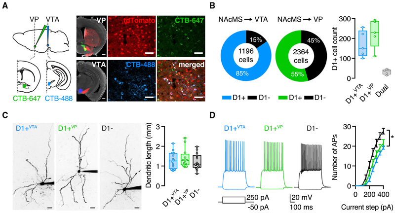
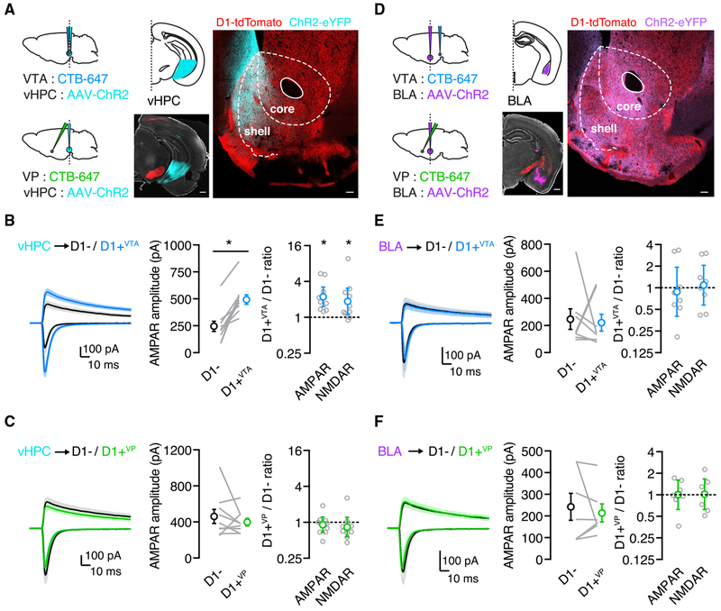
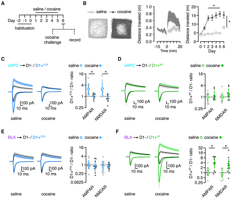
We examine synaptic connectivity and cocaine-evoked plasticity at specific networks within the nucleus accumbens (NAc). We identify distinct subpopulations of D1+ medium spiny neurons (MSNs) that project to either the ventral pallidum (D1+VP) or the ventral tegmental area (D1+VTA). We show that inputs from the ventral hippocampus (vHPC), but not the basolateral amygdala (BLA), are initially biased onto D1+VTA MSNs. However, repeated cocaine exposure eliminates the bias of vHPC inputs onto D1+VTA MSNs, while strengthening BLA inputs onto D1+VP MSNs. Our results reveal that connectivity and plasticity depend on the specific inputs and outputs of D1+ MSNs and highlight the complexity of cocaine-evoked circuit level adaptations in the NAc.

Keywords: cocaine sensitization; medium spiny neuron; nucleus accumbens; spiny projection neuron; synaptic plasticity.


Conflict of interest statement

DECLARATION OF INTERESTS

The authors declare no competing interests.

Figures

Figure 1.
Figure 1.. D1+ MSNs Projecting to VP and VTA Are Distinct Cell Populations
(A) Left: schematic and representative images for injections of CTB-647 into the VP (green) and CTB-488 into the VTA (blue) of D1-tdTomato mice (red) (scale bar, 500 μm). Right: confocal images of retrogradely labeled neurons in the NAcMS (arrows indicate dual-labeled MSNs; scale bar, 100 μm). (B) Left: quantification of CTB and tdTomato overlap in the NAcMS (total cell count of three slices per mouse, five mice). Right: summary of the number of D1+VTA, D1+VP and dual-projecting MSNs in the NAcMS (average number of labeled cells of three slices per mouse, five mice). (C) Two-photon images of D1+VTA, D1+VP, and D1− MSNs (left) (scale bar, 20 μm) and quantification of total dendritic length for all reconstructed cells (right), showing no differences. (D) Examples of physiological responses of D1+VTA, D1+VP, and D1− MSNs to current injections of 250 and −50 pA. Right: summary of current step-evoked firing, showing similar number of action potentials (APs) for D1+VTA and D1+VP MSNs. Box-and-whisker plots represent median and minimum to maximum. F-I curve is presented as mean ± SEM. *p < 0.05. See also Figure S1.
Figure 2.
Figure 2.. vHPC Inputs Are Stronger onto D1+VTA Cells in Naive Mice
(A) Left: schematic for injections of AAV-ChR2-eYFP into the vHPC and CTB-647 into the VP or the VTA. Right: representative image of viral expression in the vHPC (scale bar, 500 μm) and of dense vHPC axon labeling in the NAcMS (scale bar, 100 μm). (B) Left: vHPC inputs evoke larger EPSCs at D1+VTA MSNs compared with neighboring D1− MSNs. Middle: summary of the absolute amplitude of vHPC-evoked AMPAR EPSCs at D1− and D1+VTA MSNs, where lines indicate pairs of recorded neurons. Right: summary of D1+VTA/D1− amplitude ratios for AMPAR EPSCs at −70 mV and NMDAR EPSCs at +40 mV. (C) Similar to (B), showing no bias of vHPC inputs onto pairs of D1+VP and D1− MSNs. (D) Left: schematic for injections of AAV-ChR2-eYFP into the BLA and CTB-647 into the VP or the VTA. Right: representative image of viral expression in the BLA (scale bar, 500 μm) and of BLA axon labeling in the NAcMS (scale bar, 100 μm). (E) Left: there is no bias of BLA-evoked EPSCs at D1+VTA MSNs compared to neighboring D1− MSNs. Middle: summary of the absolute amplitude of BLA-evoked AMPAR EPSCs at D1− and D1+VTA MSNs, where lines indicate pairs of recorded neurons. Right: summary of D1+VTA/D1− amplitude ratios for AMPAR EPSCs at −70 mV and NMDAR EPSCs at +40 mV. (F) Similar to (E) showing no bias of BLA-evoked EPSCs at D1+VP MSNs compared to neighboring D1− MSNs. Average traces are presented as mean ± SEM. Ratio data are presented as geometric mean with 95% CI on logarithmic axes. *p < 0.05. See also Figure S1.
Figure 3.
Figure 3.. Cocaine-Induced Plasticity Depends on Projection Target
(A) Sensitization protocol, with 3 days of habituation, 5 days of either cocaine or saline injections, and 1 day of withdrawal, followed by either cocaine challenge to assess behavior or slice recordings to study synaptic physiology. (B) Left: typical movement of saline and cocaine mice to a challenge dose of cocaine on day 6 of the protocol. Middle: summary of distance traveled in the cocaine challenge session. Right: summary of distance traveled per experiment day. (C) Left: vHPC inputs evoke larger EPSCs at D1+VTA MSNs in saline mice, but this bias is abolished in cocaine mice. Right: summary of D1+VTA/D1− amplitude ratios. (D) Similar to (C), showing equivalent vHPC-evoked EPSCs at D1+VP and D1− MSNs in both saline and cocaine mice. (E) Left: BLA-evoked EPSCs are similar at D1+VTA and D1− MSNs in saline and cocaine mice. Right: summary of D1+VTA/D1− amplitude ratios. (F) Similar to (E) but showing a large increase in EPSCs at D1+VP MSNs in cocaine mice. Average traces are presented as mean ± SEM. Ratio data are presented as geometric mean with 95% CI on logarithmic axes. *p < 0.05. See also Figure S3.

References

    1. Ade KK, Wan Y, Chen M, Gloss B, and Calakos N (2011). An improved BAC transgenic fluorescent reporter line for sensitive and specific identification of striatonigral medium spiny neurons. Front. Syst. Neurosci 5, 32. - PMC - PubMed
    1. Al-Hasani R, McCall JG, Shin G, Gomez AM, Schmitz GP, Bernardi JM, Pyo C-O, Park SI, Marcinkiewcz CM, Crowley NA, et al. (2015). Distinct subpopulations of nucleus accumbens dynorphin neurons drive aversion and reward. Neuron 87, 1063–1077. - PMC - PubMed
    1. Ambroggi F, Ishikawa A, Fields HL, and Nicola SM (2008). Basolateral amygdala neurons facilitate reward-seeking behavior by exciting nucleus accumbens neurons. Neuron 59, 648–661. - PMC - PubMed
    1. Bock R, Shin JH, Kaplan AR, Dobi A, Markey E, Kramer PF, Gremel CM, Christensen CH, Adrover MF, and Alvarez VA (2013). Strengthening the accumbal indirect pathway promotes resilience to compulsive cocaine use. Nat. Neurosci 16, 632–638. - PMC - PubMed
    1. Bocklisch C, Pascoli V, Wong JCY, House DRC, Yvon C, de Roo M, Tan KR, and Lüscher C (2013). Cocaine disinhibits dopamine neurons by potentiation of GABA transmission in the ventral tegmental area. Science 341, 1521–1525. - PubMed

Publication types

LinkOut - more resources