. 2019 Aug 27;28(9):2397-2412.e4.
doi: 10.1016/j.celrep.2019.07.072.

VE-Cadherin Is Required for Lymphatic Valve Formation and Maintenance

Affiliations

VE-Cadherin Is Required for Lymphatic Valve Formation and Maintenance

Ying Yang et al. Cell Rep. .

Abstract

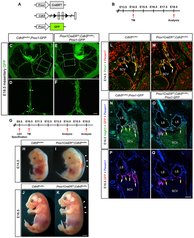
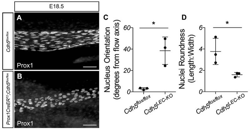
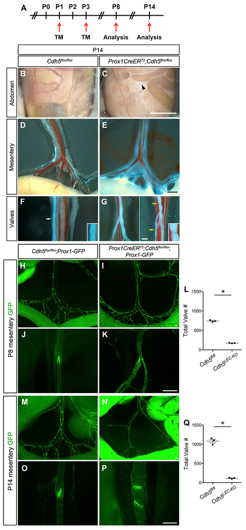
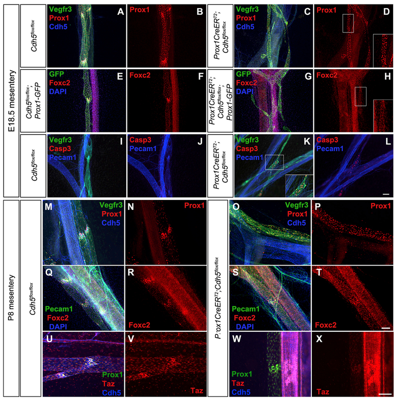
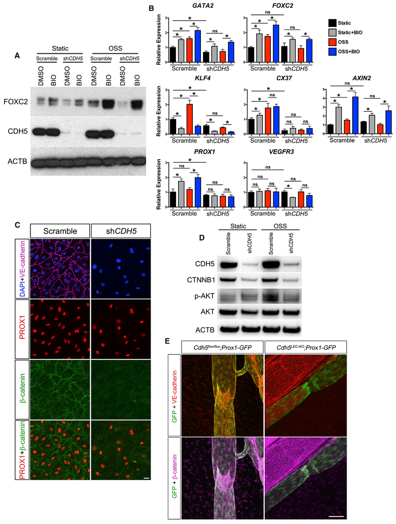
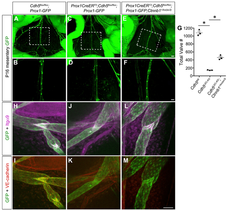
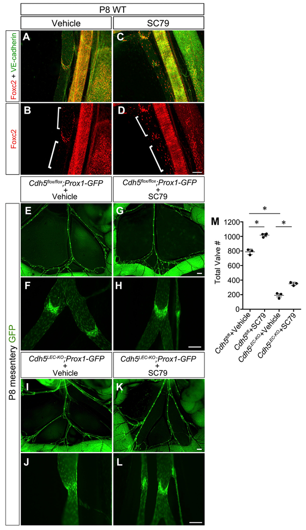
The lymphatic vasculature requires intraluminal valves to maintain forward lymph flow. Lymphatic valves form and are constantly maintained by oscillatory fluid flow throughout life, yet the earliest steps of how lymphatic endothelial cells are able to respond to fluid shear stress remain unknown. Here, we show that the adherens junction protein VE-cadherin is required for the upregulation of valve-specific transcription factors. Conditional deletion of VE-cadherin in vivo prevented valve formation in the embryo and caused postnatal regression of nearly all lymphatic valves in multiple tissues. Since VE-cadherin is known to signal through β-catenin and the VEGFR/AKT pathway, each pathway was probed. Expression of a constitutively active β-catenin mutant or direct pharmacologic activation of AKT in vivo significantly rescued valve regression in the VE-cadherin-deficient lymphatic vessels. In conclusion, VE-cadherin-dependent signaling is required for lymphatic valve formation and maintenance and therapies to augment downstream pathways hold potential to treat lymphedema in patients.

Keywords: Akt; mechanotransduction; shear stress; β-catenin.


Conflict of interest statement

DECLARATION OF INTERESTS

The authors declare no competing interests.

Figures

Figure 1.
Figure 1.. Embryonic Deletion of VE-Cadherin Causes Loss of Lymphatic Valves
(A) Alleles used to delete VE-cadherin from lymphatic endothelium while simultaneously enabling the visualization of Prox1-expressing cells. (B) Schematic of the tamoxifen schedule used to delete VE-cadherin from the embryonic mesentery. TM, tamoxifen. (C–F) Direct fluorescence imaging of GFP (green) of freshly dissected mesenteries from E18.5 Cdh5flox/flox (C and D) and Prox1CreERT2;Cdh5flox/flox (E and F) embryos. (D and F) High-magnification images of the areas outlined by the white dashed boxes in (C) and (E), respectively. White arrows indicate lymphatic valves. Images are representative of n = 3 or more mesenteries per genotype. (See also Figure S2 for loss of lymphatic valves in embryonic back skin and axillary regions.) (G) Tamoxifen schedule used for early embryonic deletion of VE-cadherin. TM, tamoxifen. (H and I) E14.5 control (H) and VE-cadherinLEC-KO (I) embryos. White arrowheads denote severe edema. (J and K) E16.5 control (J) and VE-cadherinLEC-KO (K) embryos. White arrowheads denote edema. (L and M) Immunostaining of frontal sections of E14.5 control (L) and VE-cadherinLEC-KO (M) embryos for PROX1 (green) and PECAM1 (red). LS, lymph sac; SCV, subclavian vein; IJV, internal jugular vein. (N and O) Immunostaining of frontal sections of E16.5 control (N) and VE-cadherinLEC-KO (O) embryos for VEGFR3 (green), GFP (red), and PECAM1 (blue). White arrows indicate lymphovenous valves while yellow arrows indicate venous valves only. (P and Q) Immunostaining of frontal sections of E16.5 control (P) and VE-cadherinLEC-KO (Q) embryos as in (N) and (O), but omitting the green channel to show Prox1-GFP expression (red channel). Scale bars are 200 μm in (C)–(F); 2 μm in (H)–(K); and 100 μm for (L)–(Q).
Figure 2.
Figure 2.. VE-Cadherin Is Required for Lymphatic Endothelial Cell Alignment with Flow in the Embryo
(A and B) E18.5 mesenteries from control (A) and VE-cadherinLE-KO (B) embryos immunostained for PROX1. (C) Nucleus orientation was measured using NIH FIJI software. The flow axis was defined as 0° and the absolute values of the angles were reported. (D) Nucleus roundness was measured as the length-to-width ratio. All values are means ± SEM of n = 3 independent experiments. *p < 0.05. Scale bars are 50 μm.
Figure 3.
Figure 3.. Postnatal Loss of VE-Cadherin Leads to Chylous Ascites and Loss of Valves
(A) Tamoxifen schedule used for postnatal deletion of VE-cadherin. TM, tamoxifen. (B and C) Chylous ascites fluid of VE-cadherinLEC-KO mice (C, black arrowhead) compared to control (B). (D and E) Lymph leakage from mesenteric lymphatic collecting vessels of VE-cadherinLEC-KO mesentery (E) compared to control (D). (F and G) Valve structures in the lymphatic collecting vessels of control (F, white arrow) and VE-cadherinLEC-KO (G, yellow arrows) mesenteries. Insets are zoomed-in views of the valves indicated by the arrows. (H and I) In situ fluorescence imaging of the mesenteric lymphatic vasculature at P8. Images are of control (H) and VE-cadherinLEC-KO (I) tissues expressing the Prox1-GFP reporter (green) at low magnification. (J and K) High-magnification images of P8 lymphatic valves of control (J) and VE-cadherinLEC-KO (K) mesenteries. (L) The total number of lymphatic valves in P8 control and VE-cadherinLEC-KO mesenteries. (M and N) In situ fluorescence imaging of the mesenteric lymphatic vasculature at P14 expressing the Prox1-GFP at low magnification for control (M) and VE-cadherinLEC-KO (N). (O and P) High-magnification images of P14 lymphatic valves of control (O) and VE-cadherinLEC-KO (P) mesenteries. (Q) The total number of lymphatic valves in the P14 mouse mesentery of control and VE-cadherinLEC-KO animals. Unpaired Student’s t tests were performed to compare the total valve number between control and VE-cadherinLEC-KO tissues at P8 and P14 (n = 3 per genotype per stage). All values are means ± SEM. *p < 0.05. Scale bars are 5 mm in (B) and (C); 500 μm in (D) and (E); 250 μm in (F) and (G); and 200 μm in (H)–(P).
Figure 4.
Figure 4.. VE-Cadherin-Deficient Lymphatic Valve Endothelial Cells Fail to Upregulate Prox1 and Foxc2 and Undergo Apoptosis
(A and C) Whole mount immunostaining of mesenteries collected from E18.5 control (A) and VE-cadherinLEC-KO (C) embryos for VEGFR3 (green), PROX1 (red), and VE-cadherin (blue). (B and D) The same images as in (A) and (C) showing only PROX1 (red) in control (B) and VE-cadherinLEC-KO (D). (E-H) Whole mount immunostaining of mesenteries collected from E18.5 control (E) and VE-cadherinLEC-KO (G) embryos expressing the Prox1-GFP reporter for GFP (green), FOXC2 (red), and 2-(4-amidinophenyl)-1H-indole-6-carboxamidine (DAPI; blue). Images of only FOXC2 (red) are shown for control (F) and VE-cadherinLEC-KO (H). (I–L) Whole mount immunostaining of mesenteries collected from E18.5 control (I) and VE-cadherinLEC-KO (K) embryos for VEGFR3 (green), Casp3 (red), and PECAM1 (blue). Images without VEGFR3 are shown for control (J) and VE-cadherinLEC-KO (L). (M and O) Whole mount immunostaining of mesenteries collected from P8 control (M) and VE-cadherinLEC-KO (O) pups for VEGFR3 (green), PROX1 (red), and VE-cadherin (blue). (N and P) The same images as in (M) and (O) showing only PROX1 (red) for control (N) and VE-cadherinLEC-KO (P). (Q–T) Whole mount immunostaining of mesenteries collected from P8 control (Q) and VE-cadherinLEC-KO (S) pups for PECAM1 (green), FOXC2 (red), and DAPI (blue). Images of only FOXC2 (red) are shown for control (R) and VE-cadherinLEC-KO (T). (U–X) Whole mount immunostaining of mesenteries collected from P8 control (U) and VE-cadherinLEC-KO (W) pups for PROX1 (green), TAZ (red), and VE-cadherin (blue). Images of only TAZ (red) are shown for control (V) and VE-cadherinLEC-KO (X). Insets at the lower right of some panels are the expanded views of the areas outlined by white boxes. Scale bars are 100 μm.
Figure 5.
Figure 5.. VE-Cadherin-Dependent Signaling Is Required for Transcription Factor Upregulation in Response to Oscillatory Flow In Vitro
(A) Western blot of FOXC2 and CDH5 in human dermal lymphatic endothelial cells (hdLECs) cultured for 48 h in the absence or presence of oscillatory flow with or without a targeted shRNA against CDH5. Cells were treated with vehicle or with a GSK3-β antagonist (BIO). (B) qRT-PCR was performed for the indicated genes using hdLECs cultured under no flow (black bars) or oscillatory flow (red bars), transfected with a control scramble or shRNA against CDH5 (as indicated), and treated with vehicle or BIO (gray and blue bars). All values are means ± SEM of n = 3 experiments. One-way ANOVA was performed with Sidak’s post hoc test for multiple comparisons (*p < 0.05). (C) hdLECs cultured under no-flow (static) conditions and immunostained for DAPI (blue), VE-cadherin (violet), PROX1 (red), and β-catenin (green) after infection with a scramble or shRNA targeting CDH5 for 48 h. (D) Western blot for β-catenin, pAKT, and total AKT in hdLECs under static or OSS conditions (for 5 min) treated with scramble or shRNA against CDH5. (E) Immunostaining for Prox1-GFP (green), VE-cadherin (red), and β-catenin (violet) in control and VE-cadherinLEC-KO lymphatic vessels. Scale bars are 50 μm in (C) and 100 μm in (E).
Figure 6.
Figure 6.. Constitutively Active β-Catenin Signaling Partially Rescues Valve Regression in the Absence of VE-Cadherin
(A–F) Direct fluorescence imaging of Prox1-GFP (green) of freshly dissected mesenteries from P16 Cdh5fl/fl, VE-cadherinLEC-KO, and VE-cadherinLEC-KO;Ctnnb1+/lox(ex3) pups at low (A, C, and E) and high (B, D, and F) magnification. (G) Quantification of the total number of valves in the mesenteries from each genotype. All values are means ± SEM of n = 3 littermates per genotype. (H–M) Confocal imaging of lymphatic valves stained for Integrin-α9, GFP, and VE-cadherin from control (H and I), VE-cadherinLEC-KO (J and K), or rescue conditions(L and M) to show valve morphology. One-way ANOVA with Tukey’s post hoc test was performed for multiple comparisons (*p < 0.05). Scale bars are 500 μm in (A), (C), and (E); 200 μm in (B), (D), and(F); and 50 μm in (H)–(M).
Figure 7.
Figure 7.. Direct Activation of Akt Partially Restores Valve Maintenance in the Absence of VE-Cadherin
(A-D) P8 wild-type lymphatic vessels from mice treated with vehicle (DMSO; A and B) or the small molecule AKT activator SC79 (C and D) and immunostained for FOXC2 (red) and VE-cadherin (green). (E-L) Direct fluorescence imaging of Prox1-GFP (green) of freshly dissected mesenteries from P8 pups treated as indicated at low(E, G, I, and K) and high (F, H, J, and L) magnification. (M) Quantification of the total number of valves in the mesenteries from each genotype. All values are means ± SEM of n = 3 experiments per genotype. One-way ANOVA with Tukey’s post hoc test was performed for multiple comparisons (*p < 0.05). Scale bars are 100 μm for(A)-(D); 500 μm for (E), (G), (I), and (K); and 100 μm for (F), (H), (J), and (L).

References

    1. Baeyens N, and Schwartz MA (2016). Biomechanics of vascular mechano-sensation and remodeling. Mol. Biol. Cell 27, 7–11. - PMC - PubMed
    1. Baluk P, Fuxe J, Hashizume H, Romano T, Lashnits E, Butz S, Vestweber D, Corada M, Molendini C, Dejana E, and McDonald DM (2007). Functionally specialized junctions between endothelial cells of lymphatic vessels. J. Exp. Med. 204, 2349–2362. - PMC - PubMed
    1. Bazigou E, Xie S, Chen C, Weston A, Miura N, Sorokin L, Adams R, Muro AF, Sheppard D, and Makinen T (2009). Integrin-alpha9 is required for fibronectin matrix assembly during lymphatic valve morphogenesis. Dev. Cell 17, 175–186. - PMC - PubMed
    1. Bazigou E, Lyons OT, Smith A, Venn GE, Cope C, Brown NA, and Makinen T (2011). Genes regulating lymphangiogenesis control venous valve formation and maintenance in mice. J. Clin. Invest. 121, 2984–2992. - PMC - PubMed
    1. Brouillard P, Boon L, and Vikkula M (2014). Genetics of lymphatic anomalies. J. Clin. Invest. 124, 898–904. - PMC - PubMed

Publication types

MeSH terms

LinkOut - more resources