. 2019 Aug 27;7(9):293.
doi: 10.3390/microorganisms7090293.

Bifidobacterial Transfer from Mother to Child as Examined by an Animal Model

Affiliations

Bifidobacterial Transfer from Mother to Child as Examined by an Animal Model

Walter Mancino et al. Microorganisms. .

Abstract

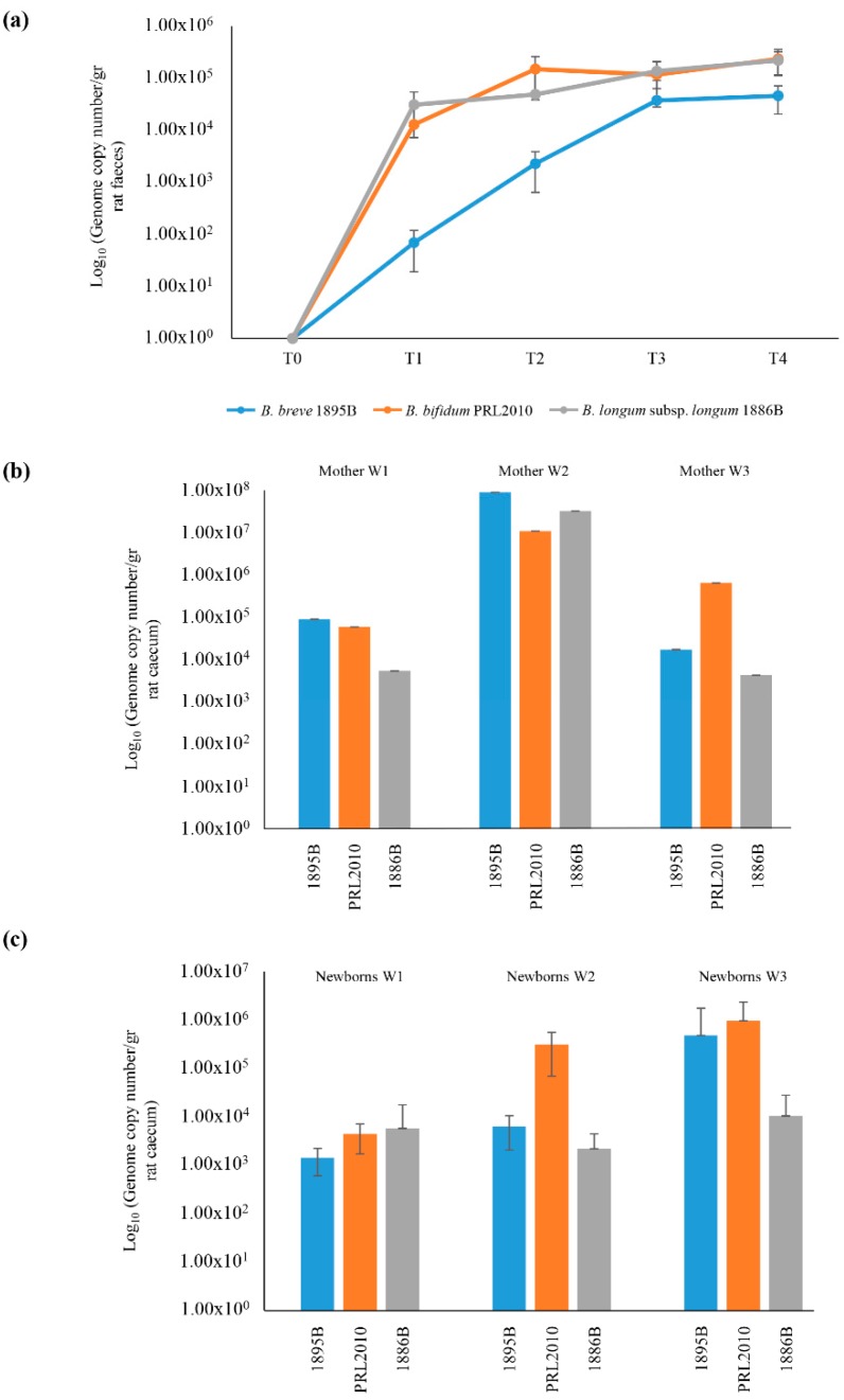
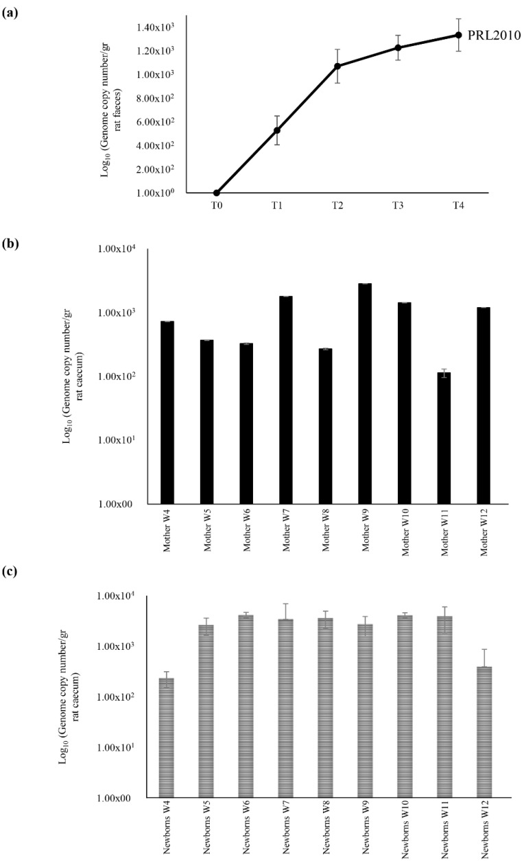
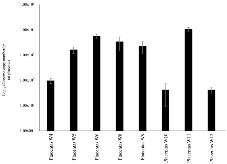
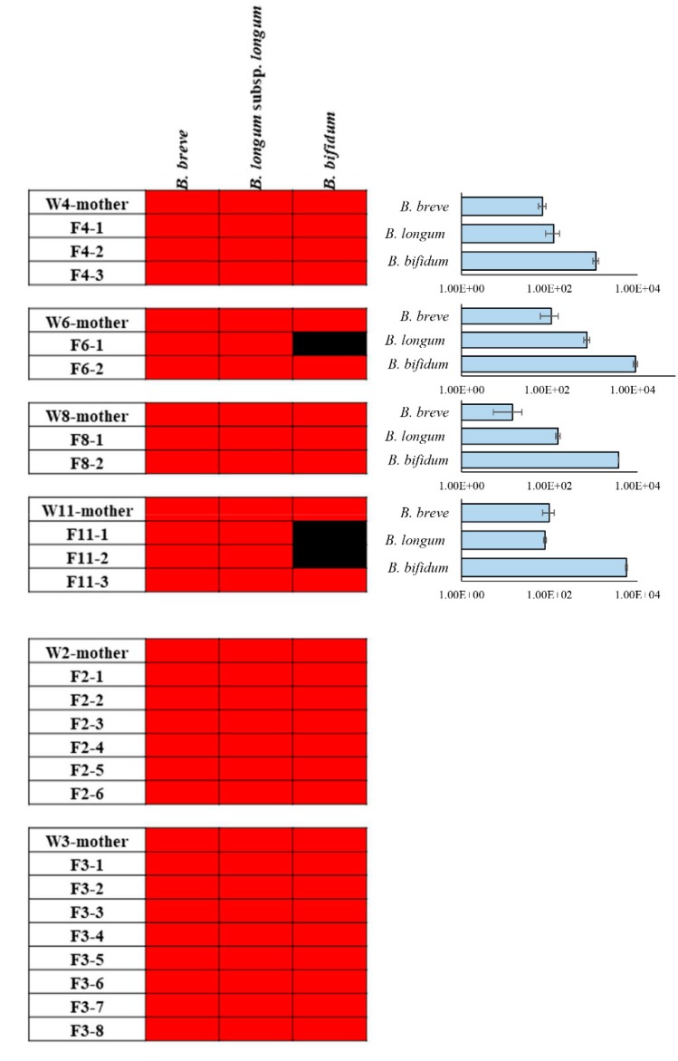
Bifidobacteria commonly constitute the most abundant group of microorganisms in the healthy infant gut. Their intestinal establishment is believed to be maternally driven, and their acquisition has even been postulated to occur during pregnancy. In the current study, we evaluated bifidobacterial mother-to infant transmission events in a rat model by means of quantitative PCR (qPCR), as well as by Internally Transcribed Spacer (ITS) bifidobacterial profiling. The occurrence of strains supplied by mothers during pregnancy to their corresponding newborns was observed and identified by analysis immediately following C-section delivery. These findings provide intriguing support for the existence of an unknown route to facilitate bifidobacterial transfer during the very early stages of life.

Keywords: bifidobacteria; infant gut microbiota; metagenomics; microbe-host coevolution.


Conflict of interest statement

The authors declare that they have no competing interests.

Figures

Figure 1
Figure 1
Schematic representation of the vertical transmission of the bifidobacterial mix in rat model treated with B. breve 1895B, B. bifidum PRL2010 and B. longum subsp. longum 1886B strains (Mix Colonization Group—MCG). Panel (a) shows the average of DNA presence of the three strains observed during the bifidobacterial administration. Each point represents the average of the log-population size ± standard deviation for three rats. Panel (b) displays the presence of B. breve 1895B, B. bifidum PRL2010 and B. longum subsp. longum 1886B in the caecum of female rats. Panel (c) exhibits bifidobacterial retrieval from puppies’ caecum. Each pillar represents the average presence for each Bifidobacterium strain ± standard deviation.
Figure 2
Figure 2
Schematic representation of vertical inheritance of B. bifidum strain PRL2010, when administered solely in a rat model (PRL2010 Group—PG). Panel (a) shows the population sizes of B. bifidum PRL2010 present in the intestine of female rats. Each point represents the average of the log-population size ± standard deviation for nine rats. Panel (b) displays the B. bifidum PRL2010 retrieval ± standard deviation for each female rat used. Panel (c) exhibits the presence of B. bifidum PRL2010 in the caecum from puppies. Each pillar represents the average colonization ± standard deviation of puppies for each female rat.
Figure 3
Figure 3
Schematic representation of the B. bifidum PRL2010 DNA load observed in placentas’ samples. This graphic displays the level of DNA belonging to B. bifidum PRL2010 present in the placenta of rats. Each pillar represents the average retrieval ± standard deviation of B. bifidum PRL2010 from the placenta of rats.
Figure 4
Figure 4
Heat map of bifidobacterial OTUs belonging to B. bifidum, B. breve and B. longum subsp. longum. Red shading represents presence, and black shading indicates absence. The pillars next to each heat map display the DNA load of these bifidobacterial species in mothers’ caeca of PG group. The x axes represent the genome copy number /gr of cecum samples.

References

    1. Ventura M., Turroni F., Motherway M.O., MacSharry J., van Sinderen D. Host-microbe interactions that facilitate gut colonization by commensal bifidobacteria. Trends Microbiol. 2012;20:467–476. doi: 10.1016/j.tim.2012.07.002. - DOI - PubMed
    1. Turroni F., Milani C., Duranti S., Ferrario C., Lugli G.A., Mancabelli L., van Sinderen D., Ventura M. Bifidobacteria and the infant gut: An example of co-evolution and natural selection. Cell. Mol. Life Sci. 2018;75:103–118. doi: 10.1007/s00018-017-2672-0. - DOI - PMC - PubMed
    1. Steed H., Macfarlane G.T., Blackett K.L., Macfarlane S., Miller M.H., Bahrami B., Dillon J.F. Bacterial translocation in cirrhosis is not caused by an abnormal small bowel gut microbiota. FEMS Immunol. Med. Microbiol. 2011;63:346–354. doi: 10.1111/j.1574-695X.2011.00857.x. - DOI - PubMed
    1. Vaishampayan P.A., Kuehl J.V., Froula J.L., Morgan J.L., Ochman H., Francino M.P. Comparative metagenomics and population dynamics of the gut microbiota in mother and infant. Genome Biol. Evol. 2010;2:53–66. doi: 10.1093/gbe/evp057. - DOI - PMC - PubMed
    1. Makino H., Kushiro A., Ishikawa E., Kubota H., Gawad A., Sakai T., Oishi K., Martin R., Ben-Amor K., Knol J., et al. Mother-to-infant transmission of intestinal bifidobacterial strains has an impact on the early development of vaginally delivered infant’s microbiota. PLoS ONE. 2013;8:e78331. doi: 10.1371/journal.pone.0078331. - DOI - PMC - PubMed