Review
. 2019 Aug 27;8(9):990.
doi: 10.3390/cells8090990.

Intraarticular Ligament Degeneration Is Interrelated with Cartilage and Bone Destruction in Osteoarthritis

Affiliations
Review

Intraarticular Ligament Degeneration Is Interrelated with Cartilage and Bone Destruction in Osteoarthritis

Gundula Schulze-Tanzil. Cells. .

Abstract

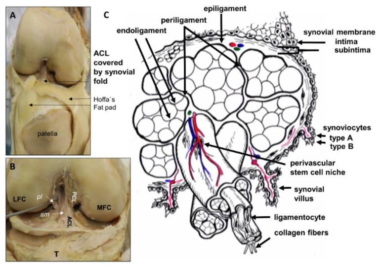
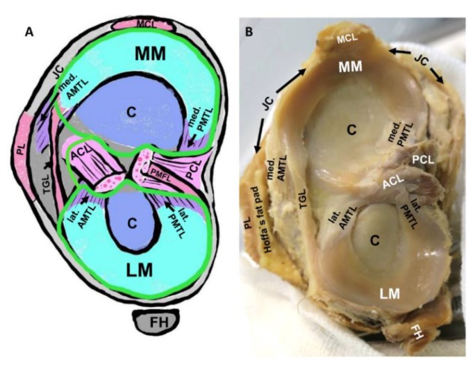
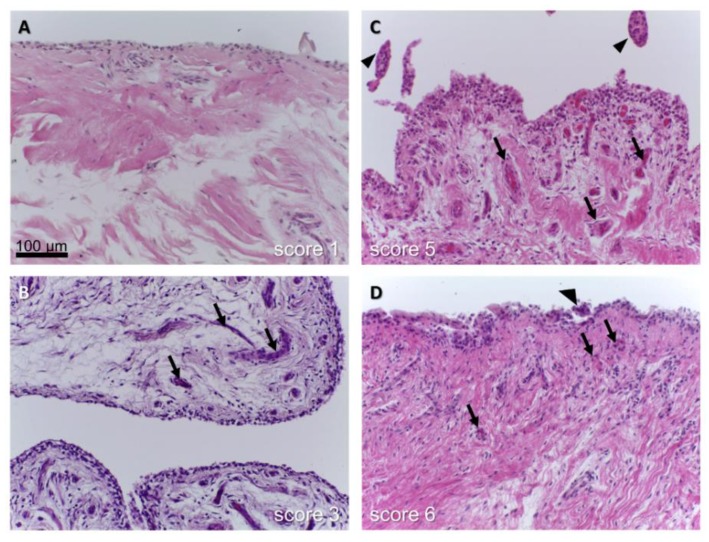
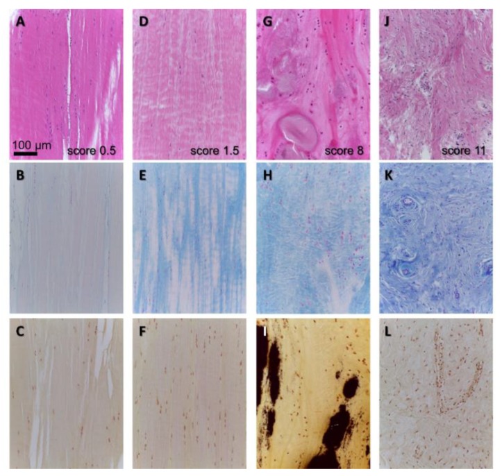
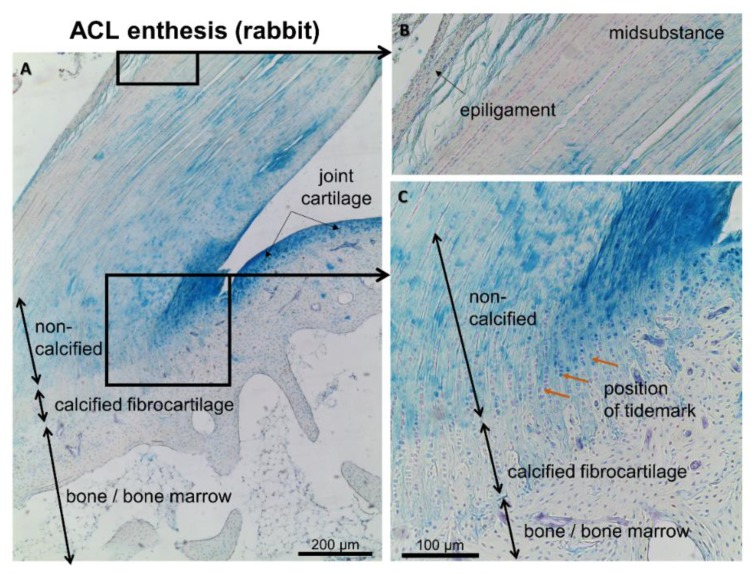
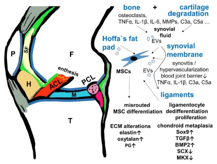
Osteoarthritis (OA) induces inflammation and degeneration of all joint components including cartilage, joint capsule, bone and bone marrow, and ligaments. Particularly intraarticular ligaments, which connect the articulating bones such as the anterior cruciate ligament (ACL) and meniscotibial ligaments, fixing the fibrocartilaginous menisci to the tibial bone, are prone to the inflamed joint milieu in OA. However, the pathogenesis of ligament degeneration on the cellular level, most likely triggered by OA associated inflammation, remains poorly understood. Hence, this review sheds light into the intimate interrelation between ligament degeneration, synovitis, joint cartilage degradation, and dysbalanced subchondral bone remodeling. Various features of ligament degeneration accompanying joint cartilage degradation have been reported including chondroid metaplasia, cyst formation, heterotopic ossification, and mucoid and fatty degenerations. The entheses of ligaments, fixing ligaments to the subchondral bone, possibly influence the localization of subchondral bone lesions. The transforming growth factor (TGF)β/bone morphogenetic (BMP) pathway could present a link between degeneration of the osteochondral unit and ligaments with misrouted stem cell differentiation as one likely reason for ligament degeneration, but less studied pathways such as complement activation could also contribute to inflammation. Facilitation of OA progression by changed biomechanics of degenerated ligaments should be addressed in more detail in the future.

Keywords: ACL; BMP signaling; chondroid metaplasia; degeneration; enthesis; ligament; meniscotibial ligaments; osteoarthritis; synovitis.


Conflict of interest statement

The author declares no conflict of interest.

Figures

Figure 1
Figure 1
Human anterior cruciate ligament (ACL) gross anatomy and scheme of its histoarchitecture. (A): Gross anatomy of the ACL (visible within the intercondylar notch) covered by the synovial membrane and neighbored to the Hoffa’s fat pad. A frontal view of the knee joint with opened joint capsule is shown. (B): The synovial membrane has been removed and the anteromedial (am) and posterolateral (pl) bundles of the ACL and the posterior cruciate ligament (PCL) attachment site became visible. (C): Scheme of the micoanatomical hierarchical structure of the ACL midsubstance. Fascicles are surrounded by the periligament, subfascicles by the endoligament. LFC, MFC: lateral and medial femoral condyles, T: Tibia.
Figure 2
Figure 2
Intraarticular meniscotibial ligaments. (A): Schematic view on the tibial plateau. (B): Human gross anatomy of the tibial plateau with menisci and meniscotibial ligaments. ACL: anterior cruciate ligament, AMTL: anterior meniscotibial ligament, C: joint cartilage, FH: fibular head (in B covered by the popliteus muscle and the lateral collateral ligament), JC: joint capsule (gray), LM: lateral meniscus, MCL: medial collateral ligament, MM: medial meniscus, PCL: posterior cruciate ligament, PMFL: posterior meniscofemoral ligament, PMTL: posterior meniscotibial ligament, TGL: transverse genicular ligament. Green line: synovial membrane.
Figure 3
Figure 3
Synovitis of the synovial membrane covering the human ACL in osteoarthritis (OA). The score reported by Berger et al., was used. (A): Unchanged synovial sheath, (B): slightly, (C,D): inflamed synovium with hypervascularization. (arrows: vessels, arrow head: villus).
Figure 4
Figure 4
Histopathological features of human ACL degeneration in OA. (A,D,G,J): HE staining to give an overview over tissue organization. (B,E,H,K): Alcian blue staining to visualize distribution of sulfated glycosaminoglycans (blue), (C,F,I,L): van Kossa staining to detect calcium deposits (black). The scoring results with the scoring system used by [11] are depicted in (A,D,G,J), (AC): nearly unchanged, (GI): chondroid metaplasia and focal calcification, (JL): ECM disintegration, hypercellularity, hypervascularization.
Figure 5
Figure 5
Enthesis of the unaffected rabbit ACL. Three different zones of the enthesis are shown using Alcian blue staining to depict sulfated glycosaminoglycan deposition (blue). (A): overview. (B): mid-substance. (C): enthesis zones.
Figure 6
Figure 6
Hypothesis of the cross-talk between cartilage/bone and ligaments in OA. A scheme of a sagittal section of the knee is shown depicting the interrelation of the joint tissues involved in OA. ACL: red, F: femur, H: Hoffa’s fad pad, brown, M: meniscus, P: patella, PCL: transected, pink, SF: synovial fluid, pale yellow, T: tibia, green: synovial membrane, gray: patellar tendon, ligament and joint capsule. BMP2: Bone morphogenetic protein, EV: extracellular vesicles, PG: proteoglycans, MKX: Mohawk, SCX: scleraxis, TGFβ: transforming growth factor β.

References

    1. Donell S. Subchondral bone remodelling in osteoarthritis. EFORT Open Rev. 2019;4:221–229. doi: 10.1302/2058-5241.4.180102. - DOI - PMC - PubMed
    1. Amini M., Nazemi S.M., Lanovaz J.L., Kontulainen S., Masri B.A., Wilson D.R., Szyszkowski W., Johnston J.D. Individual and combined effects of OA-related subchondral bone alterations on proximal tibial surface stiffness: A parametric finite element modeling study. Med. Eng. Phys. 2015;37:783–791. doi: 10.1016/j.medengphy.2015.05.011. - DOI - PubMed
    1. Ding M., Dalstra M., Linde F., Hvid I. Changes in the stiffness of the human tibial cartilage-bone complex in early-stage osteoarthrosis. Acta Orthop. Scand. 1998;69:358–362. doi: 10.3109/17453679808999047. - DOI - PubMed
    1. Nagelli C.V., Cook J.L., Kuroki K., Bozynski C., Ma R., Hewett T.E. Does Anterior Cruciate Ligament Innervation Matter for Joint Function and Development of Osteoarthritis? J. Knee Surg. 2017;30:364–371. doi: 10.1055/s-0036-1592145. - DOI - PubMed
    1. Maletius W., Messner K. Eighteen- to twenty-four-year follow-up after complete rupture of the anterior cruciate ligament. Am. J. Sports Med. 1999;27:711–717. doi: 10.1177/03635465990270060501. - DOI - PubMed

Publication types

MeSH terms