. 2019 Aug 28;7(1):230.
doi: 10.1186/s40425-019-0674-1.

Characterization of a whole blood assay for quantifying myeloid-derived suppressor cells

Affiliations

Characterization of a whole blood assay for quantifying myeloid-derived suppressor cells

Minjun C Apodaca et al. J Immunother Cancer. .

Abstract

Background: Myeloid-derived suppressor cells (MDSC) have been found to play an important role in limiting immune responses in cancer. Higher circulating MDSC levels have been associated with greater tumor burden, poorer response to immunotherapy, and poorer survival. Optimal measurement of MDSC levels could provide clinicians with a useful prognostic and/or management tool.

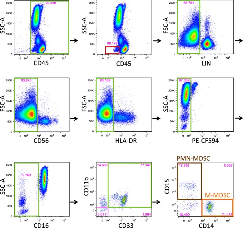
Methods: A whole blood (WB) nine color, 11 parameter flow cytometric assay was designed, utilizing fluorescently-labeled antibodies against CD45, CD3, CD19, CD20, CD56, CD16, HLA-DR, CD33, CD11b, CD14 and CD15, and BD Trucount beads for quantitation. Total MDSC were defined as CD45 + CD3-CD19-CD20-CD56-CD16-HLA-DR-CD33 + CD11b + cells, while the monocytic (M-MDSC) and polymorphonuclear subsets were defined as CD14+ or CD15+, respectively.

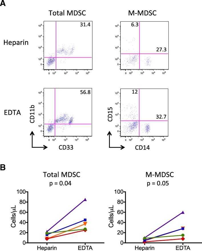
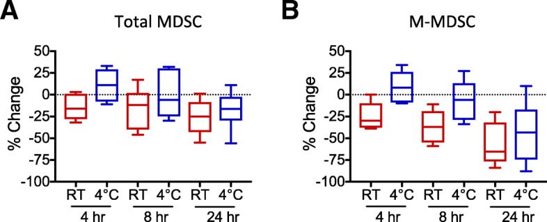
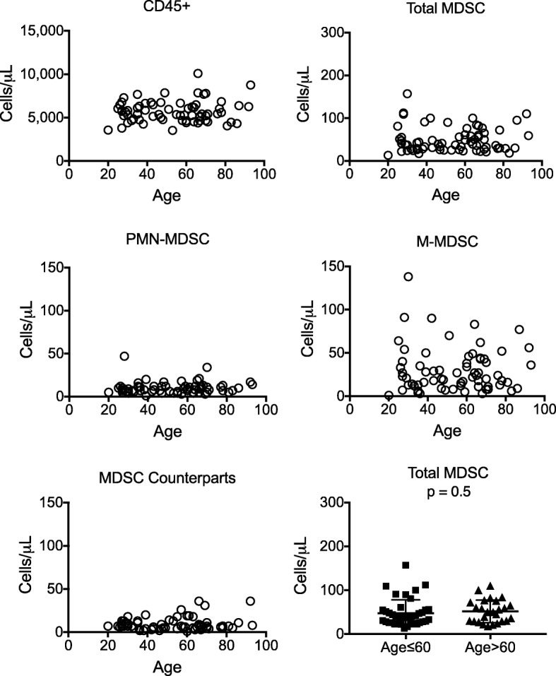
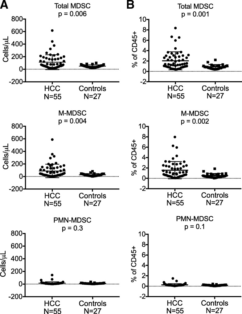
Results: A novel gating strategy was devised to eliminate granulocytes and improve consistency in gating. Several pre-analytical variables were found to significantly affect MDSC quantitation, including collection tube type and time elapsed between blood collection and testing. Total and M-MDSC levels were a mean of 63% and 73% greater, respectively, with K2EDTA compared to Na+heparin collection tubes (N = 5). In addition, time elapsed at room temperature prior to cell labeling affected MDSC quantitation; by 24 h after blood collection, total and M-MDSC levels were a mean of 26% and 57% lower compared to testing as soon as possible after collection (N = 6). Refrigeration of samples at 4 °C ameliorated time-dependent effects at both 4 and 8 h, but not 24 h after blood collection. To establish normal ranges for this assay, MDSC levels were quantified in 67 healthy subjects (30 male, 37 female) ages 20-93. No significant differences in total or M-MDSC levels were detected for ages ≤60 compared to > 60 (p = 0.5 and p = 0.8, respectively). Finally, assay results demonstrated significantly higher MDSC levels among patients with hepatocellular carcinoma (N = 55) compared to age-matched healthy controls (N = 27) for total and M-MDSC (p = 0.006 and 0.004, respectively).

Conclusions: MDSC are a heterogenous group of cells, and their quantitation in WB can be affected by a number of pre-analytical variables. Consideration of these factors, and measurement using a material type that has not been manipulated, such as whole blood, is likely to yield the most accurate results.

Keywords: Flow cytometry; Immunotherapy; Liver cancer; Myeloid-derived suppressor cells; Whole blood.


Conflict of interest statement

The authors declare they have no competing interests.

Figures

Fig. 1
Fig. 1
Gating strategy for identification of MDSC. Fresh whole blood (WB) samples (100 μL) served as the substrate for the WB flow cytometric assay. Green boxes indicate the cell populations that were selected for continued analysis. Red boxes indicate cell populations that were excluded. Initial exclusion of cellular doublets and debris, by gating on the singlets, is not shown. CD45+ cells were selected, followed by basophil exclusion, both using plots of CD45 vs SSC-A. Subsequently, T and B cells were excluded by gating on cells negative for pooled anti-CD3, anti-CD19 and anti-CD20 antibodies (lineage negative, LIN) cells. NK cells were excluded by gating on CD56 cells, and HLA-DR cells were selected. Eosinophils were excluded by gating on the PE-CF594 cell population. Neutrophils were excluded by gating on CD16 cells. Total myeloid derived suppressor cells (MDSC) were defined as CD33 + CD11b + cells. Polymorphonuclear-MDSCs (PMN-MDSC, a subset of total MDSCs, brown box) were identified by CD15+ expression, while monocytic-MDSCs (M-MDSC, a subset of total MDSCs, orange box) were identified by CD14+ expression. Early stage MDSC (e-MDSC), or counterpart MDSC in healthy individuals [7] are shown in the final plot, bottom left quadrant, as CD3/19/20/56HLA-DRCD16CD33 + CD11b + CD14CD15 cells
Fig. 2
Fig. 2
MDSC numbers in whole blood compared to PBMC, and immunosuppressive function. Total MDSC were measured using the same antibody panel in parallel using WB and PBMC from the same subjects, 2 healthy and 3 with HCC (a). Data are shown as the total MDSC percentage among CD45+ cells after imputing the number of CD45+ granulocytes to make the PBMC results comparable to the WB results. A paired t test was used to obtain the p value shown. To demonstrate suppressive activity of the MDSC identified by our assay, total MDSC (“suppressors”) were enriched as described in Materials and Methods, and cultured with autologous PBMC (“responders”) at a 1:1 ratio and stimulated with anti-CD3 and anti-CD28 beads for 4 days (right panel) (b). In addition, CD33HLA-DRCD3 cells were used as control non-suppressor cells (middle panel). CD3+ T cell proliferation was detected using intracellular Ki67 labeling for all conditions, including “responder cells only” shown in the left panel
Fig. 3
Fig. 3
Collection tube type affects MDSC quantitation. Blood samples were simultaneously collected in Na+ heparin and K2EDTA tubes and tested using the WB assay. Representative plots of total and M-MDSC populations for samples collected from the same subject in the two tube types are shown (a). Quantitative results for the total and M-MDSC populations (cells/μL) from 5 unique individuals, 2 healthy and 3 with HCC, are shown (b). Mean percent differences between K2EDTA and Na+ heparin tubes for total MDSC and M-MDSC cell counts were 63% and 73%, respectively. P-values were obtained using paired t tests
Fig. 4
Fig. 4
Temperature and time elapsed prior to testing affect MDSC quantitation. Whole blood samples from 2 healthy and 5 HCC subjects were collected in K2EDTA tubes and kept at room temperature (red box plots) or at 4 °C (blue box plots) before testing. Antibody labeling was conducted as soon as possible after blood collection, and % change in absolute numbers of total MDSC (a) and M-MDSC (b) were calculated between these baseline data and those obtained 4, 8, or 24 h after blood collection. Paired t tests were used to determine whether differences were statistically significant. At 4 h compared to baseline, mean percent change in MDSC levels for samples maintained at 4 °C versus room temperature (RT) were 9% vs − 15% change (p = 0.02) for total MDSC and 8% vs − 24% change (p = 0.009) for M-MDSC. At 8 h, mean percent changes for the 4 °C and RT samples were − 2% vs − 16% change (p = 0.06) for total MDSC and − 5% vs − 36% change (p = 0.006) for M-MDSC. Mean differences between the two conditions by 24 h were − 17% vs − 26% change (p = 0.3) for total MDSC and − 44% vs − 57% change (p = 0.4) for M-MDSC. Percent changes in MDSC counts by 24 h were lower than at 4 h (total MDSC p = 0.04 and M-MDSC p = 0.01) for samples maintained at 4 °C
Fig. 5
Fig. 5
MDSC frequencies among healthy adults. Blood samples were collected from 67 healthy subjects (30 males, 37 females) ages 20 to 93 in K2EDTA tubes. Quantitative results for CD45+, CD33 + CD11b + (total MDSC), M-MDSC (CD14+), PMN-MDSC (CD15+), MDSC counterparts (CD14CD15) from each subject are shown as individual symbols. No significant difference was found in total MDSC levels among subjects (both genders) 60 years and younger (N = 41) compared to those over the age of 60 (N = 26) (unpaired t test, p = 0.5, bottom right panel)
Fig. 6
Fig. 6
MDSC frequencies are higher among hepatocellular carcinoma (HCC) patients compared to age-matched control subjects. Whole blood samples collected from HCC patients (N = 55, mean age = 62.6, range 50–75) and healthy controls (N = 27, mean age 63.2, range 48–71) were tested using the WB MDSC assay. Results for total MDSC (CD11b + CD33+, p = 0.006) and M-MDSC (CD11b + CD33 + CD14 + CD15, p = 0.004) counts (cells/μL) among HCC patients were significantly different from healthy controls, but not for PMN-MDSC (CD11b + CD33 + CD14CD15+, p = 0.3, a). Corresponding data for total MDSC, M-MDSC, and PMN-MDSC displayed as % of CD45+ cells is also shown (b). Using absolute cell counts, twenty of the 55 HCC patients (37%) had total MDSC levels above our normal threshold of 110 cells/μL, while 19 (35%) and 5 (9%) had M-MDSC and PMN-MDSC levels above our normal thresholds of 90 cells/μL and 25 cells/μL, respectively. All p-values were obtained using unpaired t tests

References

    1. Messmer MN, Netherby CS, Banik D, Abrams SI. Tumor-induced myeloid dysfunction and its implications for cancer immunotherapy. Cancer Immunol Immunother. 2015;64(1):1–13. - PMC - PubMed
    1. Kitano S, Postow MA, Ziegler CG, Kuk D, Panageas KS, Cortez C, et al. Computational algorithm-driven evaluation of monocytic myeloid-derived suppressor cell frequency for prediction of clinical outcomes. Cancer Immunol Res. 2014;2(8):812–821. - PMC - PubMed
    1. Sade-Feldman M, Kanterman J, Klieger Y, Ish-Shalom E, Olga M, Saragovi A, et al. Clinical significance of circulating CD33+CD11b+HLA-DR- myeloid cells in patients with stage IV melanoma treated with Ipilimumab. Clin Cancer Res. 2016;22(23):5661–5672. - PubMed
    1. Weide B, Martens A, Zelba H, Stutz C, Derhovanessian E, Di Giacomo AM, et al. Myeloid-derived suppressor cells predict survival of patients with advanced melanoma: comparison with regulatory T cells and NY-ESO-1- or melan-A-specific T cells. Clin Cancer Res. 2014;20(6):1601–1609. - PubMed
    1. Tarhini AA, Edington H, Butterfield LH, Lin Y, Shuai Y, Tawbi H, et al. Immune monitoring of the circulation and the tumor microenvironment in patients with regionally advanced melanoma receiving neoadjuvant ipilimumab. PLoS One. 2014;9(2):e87705. - PMC - PubMed

Publication types

MeSH terms

Substances