

The eye in dengue fever, a rarely appreciated aspect of dengue expanded syndrome: a case report
- PMID: 31462315
- PMCID: PMC6714390
- DOI: 10.1186/s13256-019-2189-2
The eye in dengue fever, a rarely appreciated aspect of dengue expanded syndrome: a case report
Abstract
Background: Dengue fever is a mosquito-borne illness prevalent mainly in the tropics. It is feared for causing the dengue hemorrhagic spectrum of the disease leading to significant morbidity and mortality. Its rarer manifestations are categorized as the expanded dengue syndrome, and though being recognized, they are not fully appreciated and understood. The involvement of the eye in dengue fever is one such phenomenon.
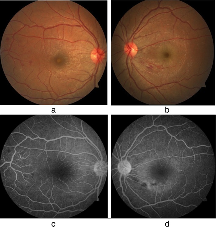
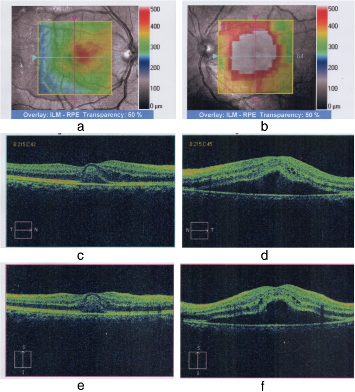
Case presentation: A 27-year-old South-Asian woman presented on day 2 of dengue fever, without capillary leakage, for further management. Despite developing hepatitis, she had an otherwise uncomplicated progression of the illness because she did not develop capillary leakage. On day 8 of the illness, she had the lowest platelet count and developed bilateral blurred vision. Examination revealed that only gross movements were detected in the left eye, and the right eye had a visual acuity of 6/9. She was diagnosed with foveolitis in the right eye and central serous chorioretinopathy in the left eye, along with hemorrhages in both eyes. These were confirmed by funduscopy, fluorescein angiography, optical coherence tomography, and macular scans. She received systemic and intravitreal steroids and was assessed regularly. After 6 months of observation, her visual acuity was 6/6 in the right eye and 6/9 in the left eye, which remained the same thereafter.
Discussion: The exact mechanism of eye involvement in dengue viral infection is poorly understood. Multiple causes have been suspected and include viral factors, immune mediation, capillary leakage, stress, and hemorrhage. Eye involvement is classically seen at the lowest platelet count and when the count begins to rise. Though symptoms are nonpathognomonic, blurring of vision is the commonest complaint, but the range of presentation is extensive and variable. Ophthalmological assessment and funduscopy are very useful in addition to advanced assessments. There is no clear consensus on management; suggestions range from conservative care to aggressive steroid therapy with immune modulation and even ophthalmological intervention. Recovery can be full or partial with a variable time scale.
Conclusion: The extensive spectrum of possible visual symptoms should prompt the clinician to suspect any visual complaint as potential dengue eye involvement. Guided studies and screening are needed to better understand the true incidence of eye involvement in dengue fever.
Keywords: Central serous chorioretinopathy; Dengue; Dengue fever; Dengue maculopathy; Dengue retinopathy; Expanded dengue syndrome; Foveolitis.
Conflict of interest statement
The authors declare that they have no competing interests.
Figures

References
-
- Gubler DJ. The global pandemic of dengue/dengue haemorrhagic fever: current status and prospects for the future. Ann Acad Med Singapore. 1998;27(2):227–234. - PubMed
-
- World Health Organization (WHO). Dengue and severe dengue. Fact sheet 117. Geneva: WHO Media Center; 2015 [updated May 2015]. http://www.who.int/mediacentre/factsheets/fs117/en/. Accessed 7 Sept 2015.
-
- World Health Organization (WHO) Regional Office for South-East Asia . Comprehensive guidelines for prevention and control of dengue and dengue haemorrhagic fever. Revised and expanded edition. Geneva: WHO; 2011.
-
- Teoh CB, Chan PL, Laude A, Chee KLC, Lim HT, Goh YK. Dengue chorioretinitis and dengue-related ophthalmic complications. https://uveitis.org/wp-content/uploads/2017/05/dengue_chorioretinitis_an.... Accessed 7 Sept 2015.
Publication types
MeSH terms
LinkOut - more resources
Full Text Sources
Medical
