. 2020 Jan;39(1):79-121.
doi: 10.1038/s41388-019-0969-1. Epub 2019 Aug 28.

LOXL2-mediated H3K4 oxidation reduces chromatin accessibility in triple-negative breast cancer cells

Affiliations

LOXL2-mediated H3K4 oxidation reduces chromatin accessibility in triple-negative breast cancer cells

J P Cebrià-Costa et al. Oncogene. 2020 Jan.

Abstract

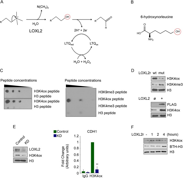
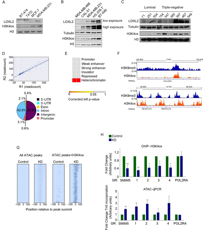
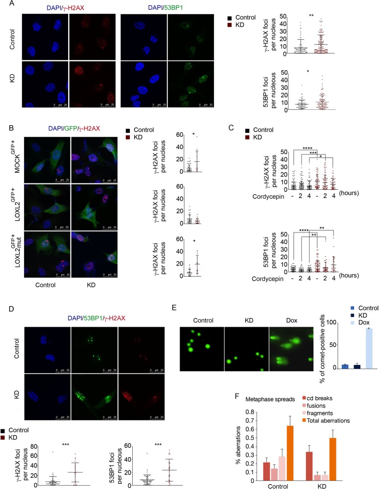
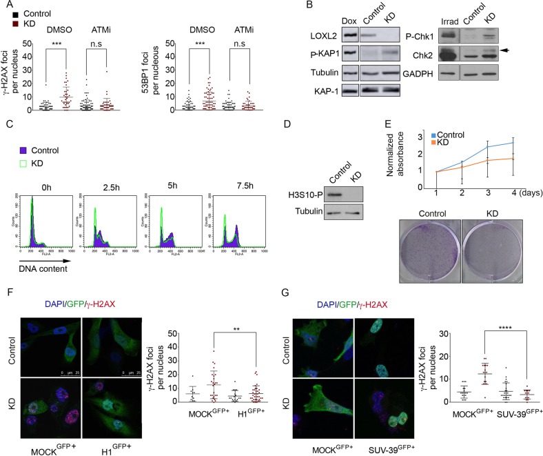
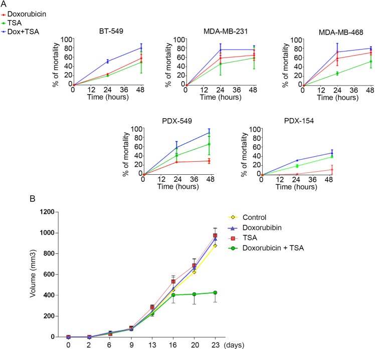
Oxidation of H3 at lysine 4 (H3K4ox) by lysyl oxidase-like 2 (LOXL2) generates an H3 modification with an unknown physiological function. We find that LOXL2 and H3K4ox are higher in triple-negative breast cancer (TNBC) cell lines and patient-derived xenografts (PDXs) than those from other breast cancer subtypes. ChIP-seq revealed that H3K4ox is located primarily in heterochromatin, where it is involved in chromatin compaction. Knocking down LOXL2 reduces H3K4ox levels and causes chromatin decompaction, resulting in a sustained activation of the DNA damage response (DDR) and increased susceptibility to anticancer agents. This critical role that LOXL2 and oxidized H3 play in chromatin compaction and DDR suggests that functionally targeting LOXL2 could be a way to sensitize TNBC cells to conventional therapy.


Conflict of interest statement

The authors declare that they have no conflict of interest.

Figures

Fig. 1
Fig. 1
Quality control of the anti-H3K4ox antibody. a Schematic representation of the LOXL2 reaction. The red circle indicates the intermediate residue that is targeted by the in-house generated anti-H3K4ox antibody. b The artificial amino acid 6-hydroxynorleucine was used in the peptide to generate the anti-H3K4ox antibody. c The anti-H3K4ox antibody was found to be specific in western blot in two replicates of dot blots of dilution series of oxidized histone H3 peptide (H3K4ox) or unmodified H3 peptide (left panel), as well as in a representative dot blot of a dilution series of H3K9me3, H3K4ox, H3K4me3, or H3 peptides (right panel). d Nucleosomes were incubated with recombinant wild-type (wt) LOXL2 or a catalytically inactive LOXL2 (mut) purified from baculovirus to detect H3K4ox/H3K4me3 levels (upper panel). Lysates of MCF-7 cells transfected with an empty vector (ø) or with LOXL2 were analyzed by western blotting, using the indicated antibodies (lower panel). e Western blot for LOXL2, H3K4ox, and total H3 from MDA-MB-231 cells infected with short hairpin RNA (shRNA) as a control, or a knockdown (KD) using a shRNA specific for LOXL2 (LOXL2 KD) (left panel). Anti-H3K4ox ChIP-PCR was used to analyze the E-cadherin gene (CDH1) promoter in MDA-MB-231 cells infected with shRNA for either control (green bar) or LOXL2 KD (blue bar). Data of qPCR amplifications were normalized to the input and to total H3 for each condition. Error bars indicate the SD from at least three independent experiments. **P < 0.01. f Western blot of H3K4ox and biotin incorporation (BTH-H3) in nucleosomes incubated with recombinant LOXL2 purified from baculovirus, after different incubation times
Fig. 2
Fig. 2
H3K4ox maps to heterochromatin and controls chromatin accessibility in TNBC cells. Western blot for the indicated antibodies in a panel of breast cancer cell lines (a), TNBC cell lines (b), or PDXs (c). d Pearson correlation between two H3K4ox sequencing replicates. Distribution of all H3K4ox ChIP-seq peaks in MDA-MB-231 cells are given, with the indicated percentages. e Contingency table of the Fisher's exact test showed the statistical overrepresentation of the H3K4ox peaks through different chromatin states. f Genome browser view of H3K4ox and H3K9me3-binding profiles at two representative heterochromatin regions. g Heatmaps show the ATAC signal in all peaks as well as in peaks that overlap with H3K4ox in LOXL2 KD or control cells. h H3K4ox ChIP-PCR validation of the selected genomic regions from the ChIP-seq from control or LOXL2 KD MDA-MB-231 cells. Data of qPCR amplification were normalized to the input and to total H3 (upper panel). ATAC-qPCR validation of the incorporation of the transposase Tn5 at the selected genomic regions from the ChIP-seq from control or LOXL2 KD MDA-MB-231 cells. Data of qPCR amplification were normalized to an unchanging genomic region (the HPRT promoter) and expressed as the fold-change relative to data obtained from control cells, which were set to 1 (lower panel). In both experiments, the RNA polymerase II (POL2RA) promoter was used as a negative control. Error bars indicate the SD from at least three independent experiments. *P < 0.05, **P < 0.01, ***P < 0.001
Fig. 3
Fig. 3
Chromatin opening activates DDR via a mechanism that requires catalytically functional LOXL2 but can be independent of DNA damage. a γ-H2AX and 53BP1 staining and foci quantification are shown by immunofluorescence with a specific antibody for γ-H2AX (left image) or for 53BP1 (right image). Dot graphs indicate the number of foci for γ-H2AX (upper graph) and 53BP1 (lower graph) per cell in control and LOXL2 KD conditions. b γ-H2AX staining and foci quantification are shown by immunofluorescence with the indicated antibody after LOXL2 reinfection. MDA-MB-231 cells were first infected with control or LOXL2 KD lentivirus and then, after puromycin selection, again with GFP (MOCKGFP+), wild-type LOXL2-IRES-GFP (LOXL2GFP+), or LOXL2mut-IRES-GFP (LOXL2mutGFP+). Cells were fixed after 24 h. Dot graphs indicate the number of γ-H2AX foci per GFP-positive cells containing MOCKGFP+ (upper graph), LOXL2GFP+ (middle graph), or LOXL2mutGFP+ (lower graph). c Dot graphs indicate the number of γ-H2AX (upper graph) and 53BP1 (lower graph) foci per cell in control and LOXL2 KD cells after treatment with 200 μM cordycepin for the indicated timepoints. d γ-H2AX and 53BP1 staining and foci quantification are shown after immunofluorescence with the indicated antibodies in non-replicative conditions. Dot graphs indicate the number of the γ-H2AX (left graph) and 53BP1 (right graph) foci in control and LOXL2 KD cells. e Representative image showing DNA damage in control or LOXL2 KD MDA-MB-231 cells, visualized by the alkaline comet assay. Cells were treated with 0.3 μM doxorubicin for 24 h. The graph shows the percentage of MDA-MB-231 positive cells. f Chromosome alterations in control and LOXL2 KD MDA-MB-231 cells. Error bars indicate the SD from at least three independent experiments. *P < 0.05, **P < 0.01, and ***P < 0.001, ****P < 0.0001
Fig. 4
Fig. 4
DDR activation is linked to chromatin decondensation in the absence of LOXL2. a Dot graphs indicate the number of foci with γ-H2AX (left graph) and 53BP1 (right graph) per cell from control and LOXL2 KD cells treated with DMSO or the ATM inhibitor KU55933. b KAP-1 phosphorylation in control or LOXL2 KD cells was analyzed by western blot. Tubulin and total KAP-1 were used as loading controls. As a positive control, MDA-MB-231 cells treated with 0.1 µM doxorubicin for 8 h (to generate DSBs) were used. Intervening lanes were removed as indicated (left panel). Chk1 and Chk2 phosphorylation in control and LOXL2 KD cells were analyzed by western blot. Chk1 phosphorylation was detected using the anti-P(S317) Chk1 antibody. For phosphorylated Chk2, a shift was detected using an anti-total Chk2 antibody. GADPH was used as a loading control. All samples were obtained under the same experimental conditions; in addition, positive control samples (irradiated fibroblasts) were run on the same gel as their corresponding control and LOXL2 KD samples. Intervening lanes in the Chk1/2 and GADPH blots were removed as indicated (right panel). c Cell cycle profile of control and LOXL2 KD cells at 0, 2.5, 5, or 7.5 h after release from a double-thymidine block. Cells were analyzed by fluorescence activated cell sorting (FACS) after propidium iodide staining. d H3S10 phosphorylation levels in control and LOXL2 KD MDA-MB-231 cells were analyzed by western blot. Tubulin was used as a loading control. e Upper panel, MTT assay in control and LOXL2 KD MDA-MB-231 cells; lower panel, colony-survival assay in control and LOXL2 KD MDA-MB-231 cells. γ-H2AX staining and foci quantification are shown by immunofluorescence with the indicated antibody after H1 (f) or SUV-39H1 reinfection (g). MDA-MB-231 cells were infected with control or LOXL2 KD lentivirus, selected with puromycin, and reinfected with GFP (MOCKGFP+), histone 1-GFP (H1GFP+) (f) or SUV-39H1-GFP (SUV-39GFP+) (g). Cells were fixed after 24 h. Dot graphs indicate the number of γ-H2AX foci per GFP-positive cell in each condition. Error bars indicate the SD from at least three independent experiments. **P < 0.01, ***P < 0.001, ****P < 0.0001
Fig. 5
Fig. 5
LOXL2 suppresses sensitivity in TNBC cells. a Cell viability was measured at different timepoints using an MTT assay of cultured different breast cancer cells and PDXs. The effects of doxorubicin, TSA, and doxorubicin plus TSA were analyzed. b Dissociated cells from PDX-549 were orthotopically implanted into NOD/SCID mice and injected intraperitoneally twice weekly with TSA (0.25 mg/kg), doxorubicin (2 mg/kg mouse weight), or doxorubicin plus TSA. Tumor volumes were measured twice a week and are given as averages. Results are given as averages of six independent tumors ± SEM

References

    1. Bannister AJ, Kouzarides T. Regulation of chromatin by histone modifications. Cell Res. 2011;21:381–95. - PMC - PubMed
    1. Williamson PR, Kagan HM. Reaction pathway of bovine aortic lysyl oxidase. J Biol Chem. 1986;261:9477–82. - PubMed
    1. Herranz N, Dave N, Millanes-Romero A, Pascual-Reguant L, Morey L, Diaz VM, et al. Lysyl oxidasE−like 2 (LOXL2) oxidizes trimethylated lysine 4 in histone H3. FEBS J. 2016;283:4263–73. - PubMed
    1. Iturbide A, Garcia de Herreros A, Peiro S. A new role for LOX and LOXL2 proteins in transcription regulation. FEBS J. 2015;282:1768–73. - PubMed
    1. Iturbide A, Pascual-Reguant L, Fargas L, Cebria JP, Alsina B, Garcia de Herreros A, et al. LOXL2 oxidizes methylated TAF10 and controls TFIID-dependent genes during neural progenitor differentiation. Mol Cell. 2015;58:755–66. - PubMed

Publication types