Review
. 2019 Oct;19(10):568-586.
doi: 10.1038/s41568-019-0183-z. Epub 2019 Aug 28.

Phagocytosis checkpoints as new targets for cancer immunotherapy

Affiliations
Review

Phagocytosis checkpoints as new targets for cancer immunotherapy

Mingye Feng et al. Nat Rev Cancer. 2019 Oct.

Abstract

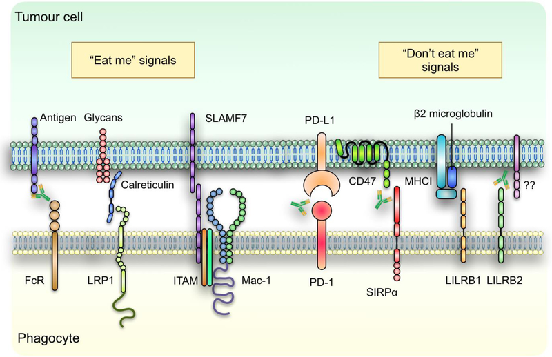
Cancer immunotherapies targeting adaptive immune checkpoints have substantially improved patient outcomes across multiple metastatic and treatment-refractory cancer types. However, emerging studies have demonstrated that innate immune checkpoints, which interfere with the detection and clearance of malignant cells through phagocytosis and suppress innate immune sensing, also have a key role in tumour-mediated immune escape and might, therefore, be potential targets for cancer immunotherapy. Indeed, preclinical studies and early clinical data have established the promise of targeting phagocytosis checkpoints, such as the CD47-signal-regulatory protein α (SIRPα) axis, either alone or in combination with other cancer therapies. In this Review, we highlight the current understanding of how cancer cells evade the immune system by disrupting phagocytic clearance and the effect of phagocytosis checkpoint blockade on induction of antitumour immune responses. Given the role of innate immune cells in priming adaptive immune responses, an improved understanding of the tumour-intrinsic processes that inhibit essential immune surveillance processes, such as phagocytosis and innate immune sensing, could pave the way for the development of highly effective combination immunotherapy strategies that modulate both innate and adaptive antitumour immune responses.


Figures

Figure 1:
Figure 1:. Phagocytosis of cancer cells is regulated by pro- and anti-phagocytic signals.
Tumour cell phagocytosis by professional phagocytes is regulated by a host of prophagocytosis (“eat me”) and antiphagocytosis (“don’t eat me) signals through receptor-ligand interactions after the cell-cell interface. Identified eat me signals expressed by tumour cells include tumour antigens which when bound by antibodies can be recognized by the Fc-receptors (FcR) on phagocytes, as well as ER chaperon protein calreticulin and SLAMF7. On the other hand, tumour cells rely on the expression of PD-L1, CD47, β2 microglobulin and yet to be identified ligands that binds to LILRB2 to inhibit their phagocytotic clearance by phagocytes. Therapeutic antibodies targeting some of these receptor-ligand interactions have been investigated as potential immunotherapy for multiple types of cancers.
Figure 2:
Figure 2:. Increased phagocytosis of tumour cells promotes activation of innate immune sensing pathways in APCs.
Following phagocytosis, degradation of tumour cells occur within phagolysosomes, which results in the recognition of tumour-derived DAMPs such as nuclear DNA (nDNA) and single stranded RNA (ssRNA) by TLRs, leading to subsequent activation of NFκB pathway. Alternatively, nucleic acids such as mitochondrial DNAs can escape from the phagosomes through a yet to be discovered mechanism into the cytosol, where they are detected by the cytosolic DNA sensor cGAS. cGAS then convert ATP and GTP into cGMP, which binds with STING to phosphorylate IRF3 and NFκB. This enables them to be translocated into the nucleus, where they act as transcription factors to promote the transcription of inflammatory cytokine genes such as type I IFNs and TNFα.
Figure 3:
Figure 3:. Increased phagocytosis of tumour cells promotes enhanced antigen cross-presentation and inflammatory cytokine release by APCs, both of which help prime effector cell responses against cancer cells.
In addition to induction of proinflammatory cytokines, phagocytosis of tumour cells also results in the release of neoantigens which are loaded onto MHCI molecules within the phagosomes. Alternative, tumour cell-derived proteins are degraded by proteasomes and shuttled back into the phagosomes or to the ER via transporter associated with antigen processing (TAP) for MHC loading. The antigen-loaded MHCs are then transported to the plasma membrane of the phagocytes, where they interact with T cell receptors (TCRs) expressed on T lymphocytes. With the help of inflammatory cytokines, the recognition of antigen-loaded MHC by TCR promotes the activation of T cells, thus enabling them to detect and eliminate tumour cells via cytotoxic responses.
Figure 4:
Figure 4:. Combination therapy with phagocytosis checkpoint blockade.
Combined modality treatment can be utilized to promote more efficient tumour cell phagocytosis and innate immune sensing pathways to produce more potent antitumor responses. For example, ionizing radiation and certain classes of chemotherapeutic compounds, can enhance the translocation of prophagocytosis signal calreticulin from the ER to the tumour cell plasma membrane, where it may synergize with CD47 blockade. Radiation and chemotherapeutic agents can also induce nuclear DNA damage, thus induce cGAS-STING mediated type I IFN responses. Finally, in addition to acting as a T cell checkpoint, PD-L1 expression on tumour cells may also inhibit their phagocytosis by macrophages. Therefore, combination therapy of CD47 and PD-L1 blockade not only should improve the phagocytic clearance of tumour cells by phagocytes, but may also lead to more potent activation of antitumour T cell immunity through enhance priming effect.

References

    1. Hodi FS et al. Improved survival with ipilimumab in patients with metastatic melanoma. N Engl J Med 363, 711–723, doi:10.1056/NEJMoa1003466 (2010). - DOI - PMC - PubMed
    1. Topalian SL et al. Safety, activity, and immune correlates of anti-PD-1 antibody in cancer. N Engl J Med 366, 2443–2454, doi:10.1056/NEJMoa1200690 (2012). - DOI - PMC - PubMed
    1. Brahmer JR et al. Safety and activity of anti-PD-L1 antibody in patients with advanced cancer. N Engl J Med 366, 2455–2465, doi:10.1056/NEJMoa1200694 (2012). - DOI - PMC - PubMed
    1. Reck M. et al. Pembrolizumab versus Chemotherapy for PD-L1-Positive Non-Small-Cell Lung Cancer. N Engl J Med 375, 1823–1833, doi:10.1056/NEJMoa1606774 (2016). - DOI - PubMed
    1. Carbone DP et al. First-Line Nivolumab in Stage IV or Recurrent Non-Small-Cell Lung Cancer. N Engl J Med 376, 2415–2426, doi:10.1056/NEJMoa1613493 (2017). - DOI - PMC - PubMed

Publication types