

Rodent Glioma Models: Intracranial Stereotactic Allografts and Xenografts
- PMID: 31462854
- PMCID: PMC6713221
- DOI: 10.1007/7657_2011_33
Rodent Glioma Models: Intracranial Stereotactic Allografts and Xenografts
Abstract
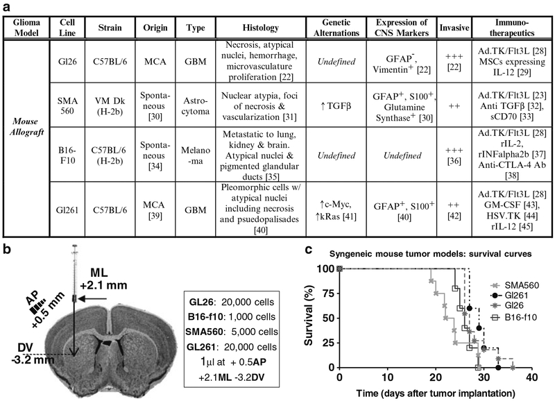
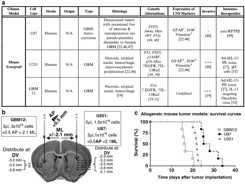
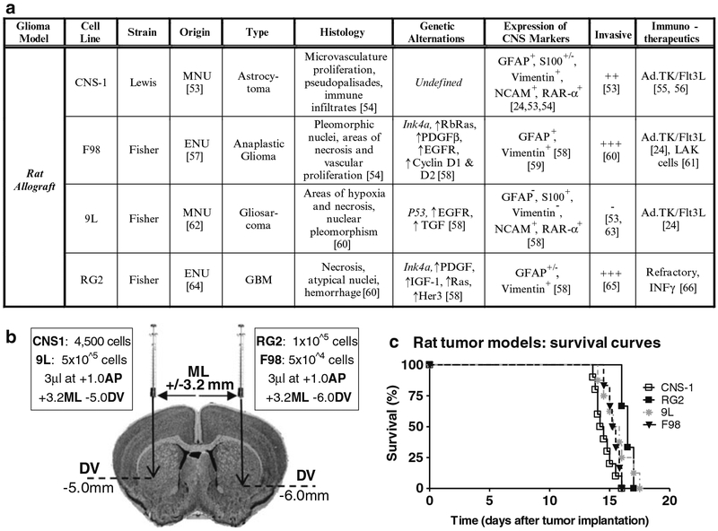
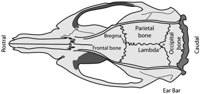
Modeling human disease in small animals has been fundamental in advancing our scientific knowledge and for the development of novel therapeutic strategies. In the case of brain cancer, implantable tumor models, both intracranial and also in the periphery, have been widely used and extensively characterized. These models can be used to better understand certain aspects of tumor biology such as growth, neovascularization, response to potential therapies, and interaction with the immune system. Brain tumors from patients as well as rodents have been cultured in vitro, in an attempt to establish permanent cell lines. Human glioma tumors have also been maintained by serial passage in the flanks of immune-deficient animals, as it has been shown that it is not feasible to continuously passage them in culture. In this chapter, we describe various gliomas that have been isolated from mice, rats, and humans and subsequently used as syngeneic or xenograft tumor models in vivo. The majority of the models that we present in this chapter arose either spontaneously or by administration of chemical carcinogens. We compare and contrast the histopathological, genetic, and invasive features of the tumor lines as well as identify novel treatment modalities that have been developed. Finally, we present the procedures for intracranial implantation of tumor cells in rodents using stereotactic surgical techniques. The use of this technique enables the generation of large numbers of animals harboring intracranial tumors with relative ease and the survival of tumor-bearing animals is highly reproducible. These characteristics make the use of these in vivo models very attractive when aiming to develop and test the effectiveness of novel anticancer therapies.
Keywords: Allograft; Brain cancer; Glioma models; Neurosurgery in rodents; Stereotactic; Tumor implantation; Xenograft.
Figures
References
-
- Burger PC, Green SB (1987) Patient age, histologic features, and length of survival in patients with glioblastoma multiforme. Cancer 59:1617–1625 - PubMed
-
- Bilzer T, Reifenberger G, Wechsler W (1989) Chemical induction of brain tumors in rats by nitrosoureas: molecular biology and neuropathology. Neurotoxicol Teratol 11:551–556 - PubMed
-
- Bulnes-Sesma S, Ullibarri-Ortiz de Zarate N, Lafuente-Sanchez JV (2006) Tumour induction by ethylnitrosourea in the central nervous system. Rev Neurol 43:733–738 - PubMed
-
- Bosch DA (1977) Short and long term effects of methyl- and ethylnitrosourea (MNU & ENU) on the developing nervous system of the rat. I. Long term effects: the induction of (multiple) gliomas. Acta Neurol Scand 55:85–105 - PubMed
Grants and funding
LinkOut - more resources
Full Text Sources