

Pulmonary echinococcosis in China
- PMID: 31463143
- PMCID: PMC6688009
- DOI: 10.21037/jtd.2019.07.31
Pulmonary echinococcosis in China
Abstract
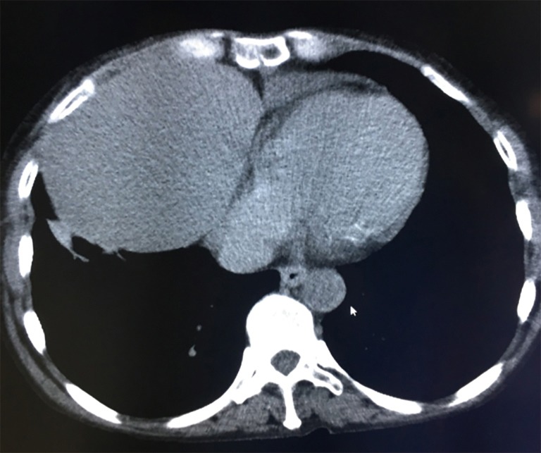
Pulmonary echinococcosis is a zoonotic parasitic disease caused by the larval stage of cestodes belonging to the genus echinococcus, which is the top killer of residents in pastoral areas and requires more attention. Prevention is the top priority; screening is another important strategy. Surgery is still the preferred treatment for pulmonary echinococcosis. Fortunately, the Chinese government has equipped each county hospital with CT machines. In the foreseeable future, the screening for pulmonary echinococcosis will begin in many areas including Tibet, which will allow us to cure early pulmonary echinococcosis in a more minimally-invasive way.
Keywords: Pulmonary; china; clinical features; diagnosis; echinococcosis; epidemiology; etiology; pathology; treatment.
Conflict of interest statement
Conflicts of Interest: The authors have no conflicts of interest to declare.
Figures


References
-
- Schad GA, Warren KS. Hookworm Disease: Current Status and New Directions. London: Taylor & Francis, 1990.
-
- Yuan ZY. Survey on cattle and sheep echinococcosis in Xinghai County of Qinghai Province. Shandong Journal of Animal Science and Veterinary Medicine 2015;36:52.
-
- Chang MH, Jin XY, Chen CY, et al. Survey on infection of echinococcosis in yaks and Tibetan sheep in Guinan County. Chinese Qinghai Journal of Animal and Veterinary Sciences 2015;45:25-6.
-
- Shu K, Qiong D, Wang L, et al. Incidence of liver echinococcosis in Naqu County of Tibet in 2014. Chinese Journal of Rural Medicine and Pharmacy 2015;22:63-4.
-
- He W, Wang Q, Huang Y, et al. Analysis of the incidence of echinococcosis in areas of Sichuan Province from 2007 to 2012. Journal of Pathogen Biology 2014;9:68-70, 91.
Publication types
LinkOut - more resources
Full Text Sources