. 2019 Aug 11:6:833-840.
doi: 10.1016/j.toxrep.2019.08.001. eCollection 2019.

Lead (Pb) exposure induces dopaminergic neurotoxicity in Caenorhabditis elegans: Involvement of the dopamine transporter

Affiliations

Lead (Pb) exposure induces dopaminergic neurotoxicity in Caenorhabditis elegans: Involvement of the dopamine transporter

Ayodele Jacob Akinyemi et al. Toxicol Rep. .

Abstract

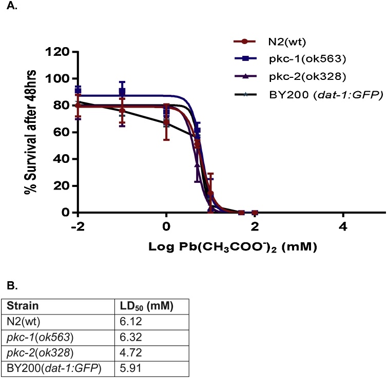
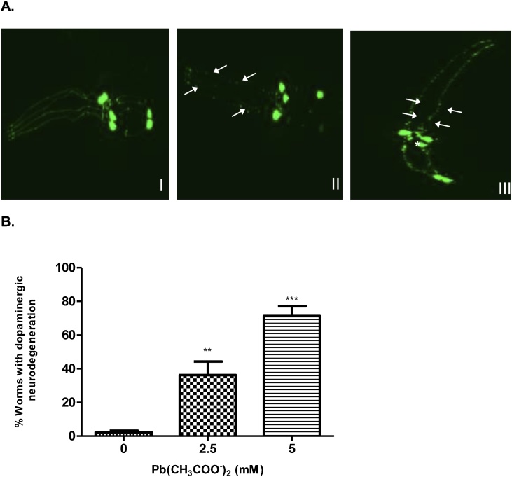
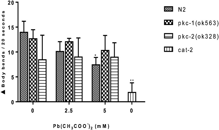
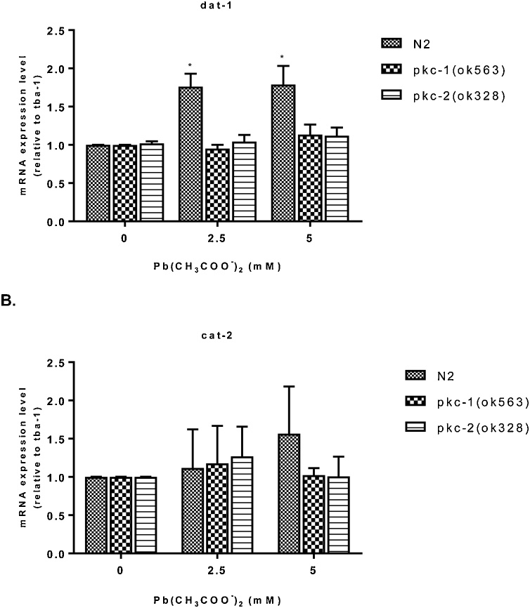
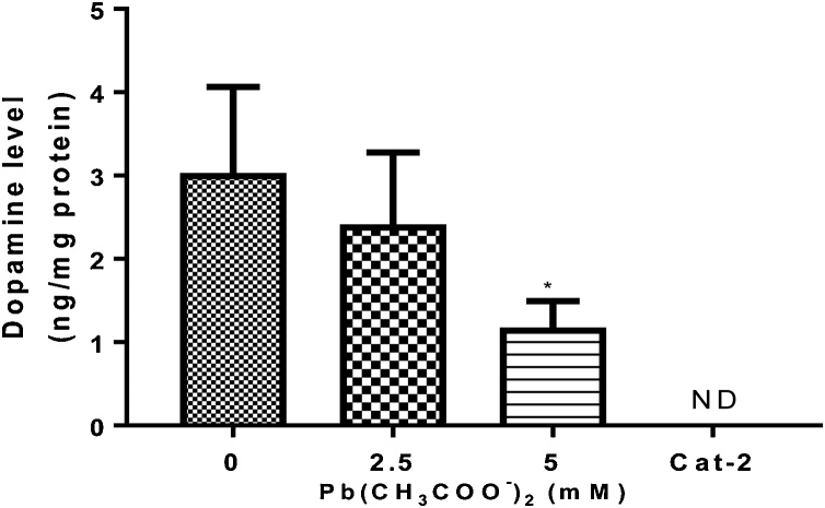
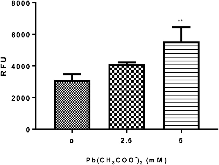
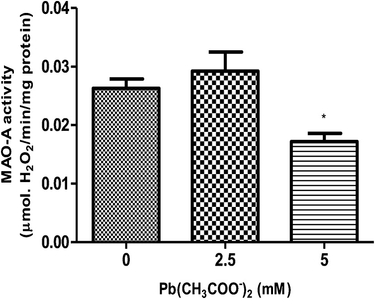
Lead (Pb) is an environmental neurotoxicant, and has been implicated in several neurological disorders of dopaminergic dysfunction; however, the molecular mechanism of its toxicity has yet to be fully understood. This study investigated the effect of Pb exposure on dopaminergic neurodegeneration and function, as well as expression level of several dopaminergic signaling genes in wild type (N2) and protein kinase C (pkc) mutant Caenorhabditis elegans. Both N2 and pkc mutant worms were exposed to Pb2+ for 1 h. Thereafter, dopaminergic (DAergic) neurodegeneration, behavior and gene expression levels were assessed. The results revealed that Pb2+ treatment affects dopaminergic cell morphology and structure in worms expressing green fluorescent protein (GFP) under a DAergic cell specific promoter. Also, there was a significant impairment in dopaminergic neuronal function as tested by basal slowing response (BSR) in wild-type, N2 worms, but no effect was observed in pkc mutant worms. Furthermore, Pb2+ exposure increased dat-1 gene expression level when compared with N2 worms, but no alteration was observed in the pkc mutant strains. LC-MS analysis revealed a significant decrease in dopamine content in worms treated with Pb2+ when compared with controls. In summary, our results revealed that Pb2+ exposure induced dopaminergic dysfunction in C. elegans by altering dat-1 gene levels, but pkc mutants showed significant resistance to Pb2+ toxicity. We conclude that PKC activation is directly involved in the neurotoxicity of Pb.

Keywords: Dopaminergic neuron; Neurotoxicant; PKC; Pb; dat-1.


Conflict of interest statement

The authors declare no conflict of interest.

Figures

Fig. 1
Fig. 1
Dose-response lethality curves of C. elegans after exposure to Pb (CH3COO)2 for 1 h. Synchronized L1 worms were exposed to increasing concentrations of Pb2+ acetate (0–50 mM) for I h. Thereafter, 30–50 worms were plated and score for lethality 48 h later. (A) Data represent the percentage of surviving worms and (B) Median lethal dose (LD50) which was calculated by non-linear regression. Sigmoidal dose-response model was used to plot the % survival curves and determine the respective LD50. Results were analyzed by two-way analysis of variance (ANOVA) followed by post hoc Tukey’s test.
Fig. 2
Fig. 2
Dopaminergic neurodegeneration in BY200 worms following Pb acetate treatment. (A) Representative confocal images of cephalic (CEP) dopaminergic neurons in head of L1 BY200 (dat-1:GFP) worms treated with Pb acetate (0, 2.5 or 5 mM), I – show intact neurons while II and III – show degenerating neurons in worms. (B) Data represent the percentage of worms with dopaminergic neurodegeneration. Results were expressed as mean ± S.E.M. (n = 6). Statistical analysis was performed by Two-way analysis of variance (ANOVA) followed by post hoc Tukey’s test. **p < 0.01,***p < 0.001 compared to control group treated with K-medium.
Fig. 3
Fig. 3
Basal slowing response (BSR) in N2 and null mutant (pkc-1 and pkc-2) worms following Pb acetate treatment for 1 h (0, 2.5 and 5 mM). BSR was carried out 48 h after Pb2+ exposure. Worms were washed off plates with S-basal and five worms were pipetted in plate’s presence or absence of OP50 E. coli. Worms were allowed to acclimatize for about 5 min. on the new plate and thereafter the body bends were counted in 20 s intervals for each worm to obtain an average. The difference in the average number of body bends in the presence and absence of food was calculated as the BSR. cat-2 mutants, with deficiency in tyrosine hydroxylase, were used as positive control. Data were expressed as mean ± S.E.M. (n = 5). Statistical analysis was performed by Two-way analysis of variance (ANOVA) followed by post hoc Tukey’s test. *p < 0.05,**p < 0.01 compared to control group treated with K-medium.
Fig. 4
Fig. 4
mRNA expression level of N2 or null mutants pkc-1 and pkc-2 worms exposed to Pb acetate for 1 h (0, 2.5 and 5 mM). (A) dat-1 expression level and (B) cat-2 expression level relative to the constitutive gene tba-1 and normalized to the N2 control group. Data were expressed as mean ± S.E.M. (n = 6). Statistical analysis was performed by Two-way analysis of variance (ANOVA) followed by post hoc Tukey’s test. *p < 0.05,**p < 0.01 compared to control group treated with K-medium.
Fig. 5
Fig. 5
Dopamine (DA) levels in N2 worms following Pb acetate treatment for 1 h (0, 2.5 and 5 mM). After 1 h treatment, DA levels were quantified by LC/MS analysis in extracts from L1 worms. Data were expressed as mean ± S.E.M. (n = 5). Statistical analysis was performed by one-way analysis of variance (ANOVA) followed by post hoc Tukey’s test. *p < 0.05 compared to control group treated with K-medium. ND – Not detected
Fig. 6
Fig. 6
Quantification of GFP fluorescence in VP596 worms expressing GFP under the gst-4 promoter following Pb acetate treatment for 1 h (0, 2.5 and 5 mM). Transgenic worms expressing GFP under the gst-4 promoter were treated with Pb2+ for 1 h (0, 2.5 and 5 mM) and GFP fluorescence was measured as an indicator of oxidative stress and SKN-1 activity. Data were expressed as mean ± S.E.M. (n = 5). Statistical analysis was performed by Two-way analysis of variance (ANOVA) followed by post hoc Tukey’s test. **p < 0.01 compared to control group treated with K-medium.
Fig. 7
Fig. 7
Monoamine oxidase (MAO-A) activity in WT N2 worms following Pb acetate treatment for 1 h (0, 2.5 and 5 mM). Data were expressed as mean ± S.E.M. (n = 5). Statistical analysis was performed by Two-way analysis of variance (ANOVA) followed by post hoc Tukey’s test. *p < 0.05 compared to control group treated with K-medium.

References

    1. Iversen S.D., Iversen L.L. Dopamine: 50 years in perspective. Trends Neurosci. 2007;30:188–193. - PubMed
    1. Kristensen A.S., Andersen J., Jorgensen T.N., Sorensen L., Eriksen J., Loland C.J., Stromgaard K., Gether U. SLC6 neurotransmitter transporters: structure, function, and regulation. Pharmacol. Rev. 2011;63:585–640. - PubMed
    1. Felton C.M., Johnson C.M. Dopamine signaling in C. elegans is mediated in part by HLH-17-dependent regulation of extracellular dopamine levels. G3 Gene. 2014;4:1081–1089. - PMC - PubMed
    1. Pramod A.B., Foster J., Carvelli L., Henry L.K. SLC6 transporters: structure, function, regulation, disease association and therapeutics. Mol. Aspects Med. 2013;34:197–219. - PMC - PubMed
    1. Chase D.L., Koelle M.R. Vol. 20. 2007. Biogenic amine neurotransmitters in C. elegans; pp. 1–15. (WormBook). Feb. - PMC - PubMed

LinkOut - more resources