Review
. 2019 Jul;8(4):456-470.
doi: 10.21037/acs.2019.07.05.

Intramural hematoma and penetrating ulcer in the descending aorta: differences and similarities

Affiliations
Review

Intramural hematoma and penetrating ulcer in the descending aorta: differences and similarities

Arturo Evangelista et al. Ann Cardiothorac Surg. 2019 Jul.

Abstract

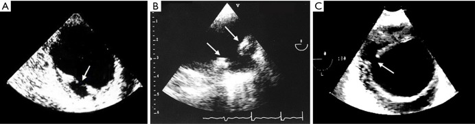
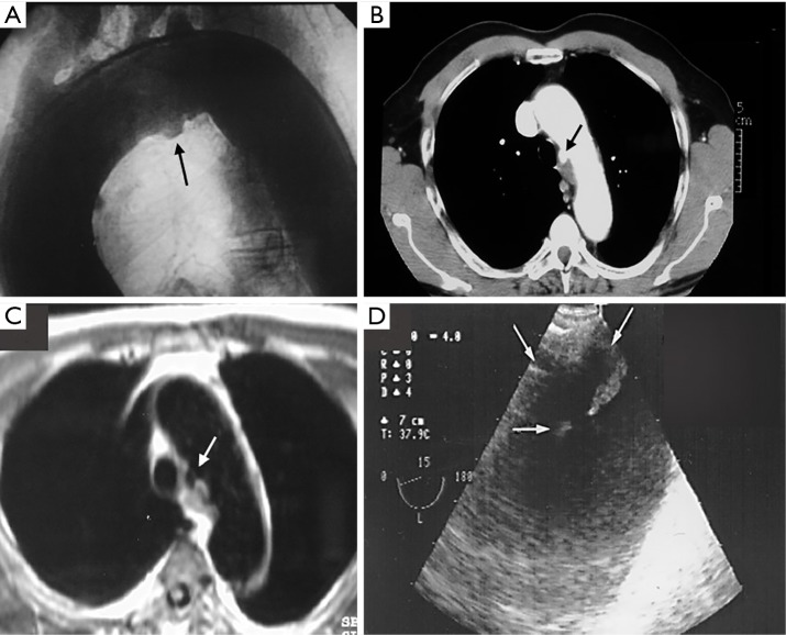
Acute aortic syndromes include a variety of overlapping clinical and anatomic diseases. Intramural hematoma (IMH), penetrating atherosclerotic ulcer (PAU), and aortic dissection can occur as isolated processes or can be found in association. All these entities are potentially life threatening, so prompt diagnosis and treatment is of paramount importance. IMH and PAU affect patients with atherosclerotic risk factors and are located in the descending aorta in 60-70% of cases. IMH diagnosis can be correctly made in most cases. Aortic ulcer is a morphologic entity which comprises several entities-the differential diagnosis includes PAU, focal intimal disruptions (FID) in the context of IMH evolution and ulcerated atherosclerotic plaque. The pathophysiologic mechanism, evolution and prognosis differ somewhat between these entities. However, most PAU are diagnosed incidentally outside the acute phase. Persistent pain despite medical treatment, hemodynamic instability, maximum aortic diameter (MAD) >55 mm, significant periaortic hemorrhage and FID in acute phase of IMH are predictors of acute-phase mortality. In these cases, TEVAR or open surgery should be considered. In non-complicated IMH or PAU, without significant aortic enlargement, strict control of cardiovascular risk factors and frequent follow-up imaging appears to be a safe management strategy.

Keywords: Intramural hematoma (IMH); acute aortic syndrome (AAS); aortic ulcer (AU); focal intimal disruption; penetrating atherosclerotic ulcer (PAU).


Conflict of interest statement

Conflicts of Interest: The authors have no conflicts of interest to declare.

Figures

Figure 1
Figure 1
Acute type B IMH. (A) CT is useful in early diagnosis, particularly, if we do not use contrast (arrow shows this high attenuation area secondary to a recent bleed); (B) CMR facilitates diagnosis by the hyperintense imaging in T2 (arrow); (C) TEE shows he intramural hematoma with mild calcification of the intima (arrow). IMH, intramural hematoma; CMR, cardiovascular magnetic resonance; TEE, transesophageal echocardiography
Figure 2
Figure 2
Tiny intimal disruptions. (A) TEE with color Doppler may be superior to any other technique for assessing the intima and demonstrating small communications (arrows) usually associated with intercostal or lumbar artery ostia; (B,C) contrast CT enhancement demonstrating a tiny intimal disruption through intimal micro-tear (black arrow), associated with an artery branch (white arrows), which disappears with IMH reabsorption. TEE, transesophageal echocardiography; IMH, intramural hematoma.
Figure 3
Figure 3
Aortic ulcers by CT: (A) penetrating atherosclerotic ulcer (arrow) in a calcified aortic arch; (B) focal intimal disruption with localized dissection (arrow); (C) angiotomography with multiplanar reconstruction (MPR) showing a large PAU in distal thoracic aorta (arrow). PAU, penetrating atherosclerotic ulcer.
Figure 4
Figure 4
Usefulness of TEE in the differential diagnosis of aortic ulcer types: (A) ulcerated plaque in descending thoracic aorta intima without involvement of the media layer of aortic wall (arrow); (B) atherosclerotic ulcerated lesion penetrating through the aortic intima into the aortic wall that progresses to the tunica media (arrows) and is diagnosed by a pouch-like protrusion into the aortic wall; (C) intramural hematoma evolution showing a localized dissection from a focal intimal disruption (FID) with an orifice diameter of 4 mm (arrow). TEE, transesophageal echocardiography.
Figure 5
Figure 5
Penetrating aortic ulcer located in the aortic arch (arrows) diagnosed by different imaging techniques: aortography (A); angio CT (B); CMR (C) and TEE (D). Only TEE identified the localized dissection (FID) from the natural evolution of intramural hematoma. CMR, cardiovascular magnetic resonance; TEE, transesophageal echocardiography; FID, focal intimal disruption.
Figure 6
Figure 6
Complicated intramural hematoma in acute phase. (A) Periaortic hematoma with large hemothorax (arrows); (B) FID with a large pseudoaneurysms (ulcer-like projection) (arrow). FID, focal intimal disruption.
Figure 7
Figure 7
Acute and chronic management pathway for type B IMH. FID, focal intimal disruption; MAD, maximum aortic diameter; IMH, intramural hematoma.
Figure 8
Figure 8
Acute and chronic management pathway for type B PAU. IMH, intramural hematoma; PAU, penetrating atherosclerotic ulcer.
Figure 9
Figure 9
Initial type B IMH (A) evolving with localized dissections (asterisks) at 8 months of follow-up (B). IMH, intramural hematoma.
Figure 10
Figure 10
Evolution and management of type B IMH. (A) IMH in acute phase diagnosed by CMR (arrow); (B) significant enlargement of descending thoracic aorta during the first week of admission (arrow); (C) angiography during endovascular treatment; (D) control by angio CT one week after TEVAR procedure. IMH, intramural hematoma; CMR, cardiovascular magnetic resonance.
Video
Video
Intramural hematoma and penetrating ulcer in the descending aorta: differences and similarities.

References

    1. Gore I. Pathogenesis of dissecting aneurysm of the aorta. AMA Arch Pathol 1952;53:142-53. - PubMed
    1. Nienaber CA, von Kodolitsch Y, Petersen B, et al. Intramural hemorrhage of the thoracic aorta: diagnostic and therapeutic implications. Circulation 1995;92:1465-72. 10.1161/01.CIR.92.6.1465 - DOI - PubMed
    1. Vilacosta I, San Roman JA. Acute aortic syndrome. Heart 2001;85:365-8. 10.1136/heart.85.4.365 - DOI - PMC - PubMed
    1. O’Gara PT, DeSanctis RW. Acute aortic dissection and its variants. Circulation 1995;92:1376-8. 10.1161/01.CIR.92.6.1376 - DOI - PubMed
    1. Nienaber CA, Sievers HH. Intramural hematoma in acute aortic syndrome – More than one variant of dissection? Circulation 2002;106:284-5. 10.1161/01.CIR.0000023453.90533.82 - DOI - PubMed