. 2018 Dec 31:19:100604.
doi: 10.1016/j.bbrep.2018.12.008. eCollection 2019 Sep.

7-Ketocholesterol and cholestane-triol increase expression of SMO and LXRα signaling pathways in a human breast cancer cell line

Affiliations

7-Ketocholesterol and cholestane-triol increase expression of SMO and LXRα signaling pathways in a human breast cancer cell line

Debora Levy et al. Biochem Biophys Rep. .

Abstract

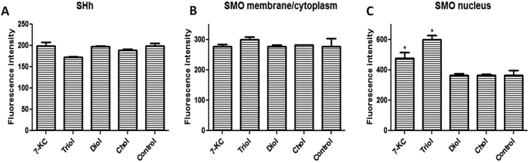
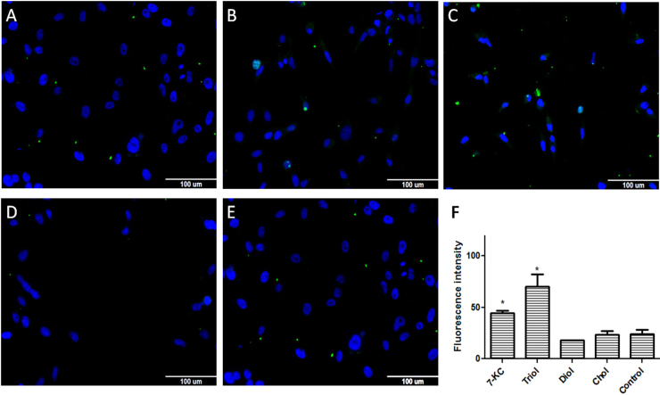
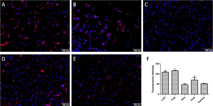
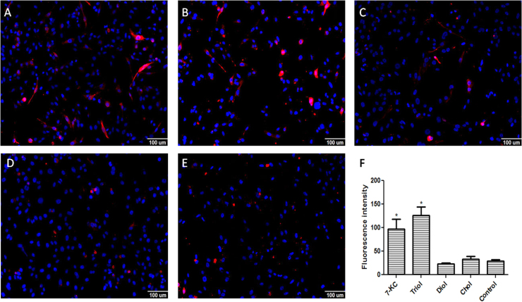
Oxysterols are 27-carbon oxidation products of cholesterol metabolism. Oxysterols possess several biological actions, including the promotion of cell death. Here, we examined the ability of 7-ketocholesterol (7-KC), cholestane-3β-5α-6β-triol (triol), and a mixture of 5α-cholestane-3β,6β-diol and 5α-cholestane-3β,6α-diol (diol) to promote cell death in a human breast cancer cell line (MDA-MB-231). We determined cell viability, after 24-h incubation with oxysterols. These oxysterols promoted apoptosis. At least part of the observed effects promoted by 7-KC and triol arose from an increase in the expression of the sonic hedgehog pathway mediator, smoothened. However, this increased expression was apparently independent of sonic hedgehog expression, which did not change. Moreover, these oxysterols led to increased expression of LXRα, which is involved in cellular cholesterol efflux, and the ATP-binding cassette transporters, ABCA1 and ABCG1. Diols did not affect these pathways. These results suggested that the sonic hedgehog and LXRα pathways might be involved in the apoptotic process promoted by 7-KC and triol.

Keywords: ABC transporters; Apoptosis; Cancer; LXRα; Oxysterol; Sonic hedgehog.


Figures

Fig. 1
Fig. 1
Immunofluorescence detection of sonic hedgehog (SHh) and smoothened (SMO) expression in MDA-MB-231 cell line after 24 h incubation with 30 µM oxysterols. A: SHh expression; B: SMO expression in the membrane/cytoplasm;C: SMO expression in the nucleus. The intensity of fluorescence was quantified with MetaXpress software. Abbreviations: 7KC: 7-ketocholesterol; triol: cholestan-3α-5β-6α-triol; diol: 5α-cholestane-3β,6β-diol/5α-cholestane-3β,6α-diol. Cholesterol was used as control. Data are presented as the mean ± SEM from three independent experiments performed in triplicate. *p < 0.005 compared to control.
Fig. 2
Fig. 2
Immunofluorescence detection of LXRα expression in the MDA-MB-231 cell line after 24 h incubation with 30 µM of oxysterols. The intensity of fluorescence was quantified with MetaXpress software. Representative images of immunofluorescence after incubations with A: 7-ketocholesterol (7KC); B: cholestan-3α-5β-6α-triol (triol); C: 5α-cholestane-3β,6β-diol/5α-cholestane-3β,6α-diol (diol); or D: cholesterol (control); E: quantification of LXRα expression. Data are presented as the mean ± SEM from three independent experiments performed in triplicate. *p < 0.005 compared to control.
Fig. 3
Fig. 3
Immunofluorescence detection of ABCA1 expression in the MDA-MB-231 cell line after 24 h incubation with 30 µM of oxysterols. The intensity of fluorescence was quantified with MetaXpress software. Representative images of immunofluorescence after incubations with A: 7-ketocholesterol (7KC); B: cholestan-3α-5β-6α-triol (triol); C: 5α-cholestane-3β,6β-diol/5α-cholestane-3β,6α-diol (diol); or D: cholesterol (control); E: quantification of ABCA1 expression. Data are presented as the mean ± SEM from three independent experiments performed in triplicate. *p < 0.005 compared to control.
Fig. 4
Fig. 4
Immunofluorescence detection of ABCG1 expression in the MDA-MB-231 cell line after 24 h incubation with 30 µM of oxysterols. The intensity of fluorescence was quantified with MetaXpress software. Representative images of immunofluorescence after incubations with A: 7-ketocholesterol (7KC); B: cholestan-3α-5β-6α-triol (triol); C: 5α-cholestane-3β,6β-diol/5α-cholestane-3β,6α-diol (diol); or D: cholesterol (control); E: quantification of ABCG1 expression. Data are presented as the mean ± SEM from three independent experiments performed in triplicate. *p < 0.005 compared to control.

References

    1. Howe V., Sharpe L.J., Alexopoulos S.J. Cholesterol homeostasis: how do cells sense sterol excess? Chem. Phys. Lipids. 2016;199:170–178. - PubMed
    1. Jakobsson T., Treuter E., Gustafsson J.A. Liver X receptor biology and pharmacology: new pathways, challenges and opportunities. Trends Pharmacol. Sci. 2012;33:394–404. - PubMed
    1. Carvalho J.F., Silva M.M., Moreira J.N. Selective cytotoxicity of oxysterols through structural modulation on rings A and B. Synthesis, in vitro evaluation, and SAR. J. Med. Chem. 2011;54:6375–6393. - PubMed
    1. Kulig W., Cwiklik L., Jurkiewicz P. Cholesterol oxidation products and their biological importance. Chem. Phys. Lipids. 2016;199:144–160. - PubMed
    1. Zerbinati C., Iuliano L. Cholesterol and related sterols autoxidation. Free Radic. Biol. Med. 2017;111:151–155. - PubMed