. 2020 Jan;9(1):106-119.
doi: 10.1002/sctm.19-0136. Epub 2019 Aug 29.

Radiation mitigation of the intestinal acute radiation injury in mice by 1-[(4-nitrophenyl)sulfonyl]-4-phenylpiperazine

Affiliations

Radiation mitigation of the intestinal acute radiation injury in mice by 1-[(4-nitrophenyl)sulfonyl]-4-phenylpiperazine

Sara Duhachek-Muggy et al. Stem Cells Transl Med. 2020 Jan.

Abstract

The objective of the study was to identify the mechanism of action for a radiation mitigator of the gastrointestinal (GI) acute radiation syndrome (ARS), identified in an unbiased high-throughput screen. We used mice irradiated with a lethal dose of radiation and treated with daily injections of the radiation mitigator 1-[(4-nitrophenyl)sulfonyl]-4-phenylpiperazine to study its effects on key pathways involved in intestinal stem cell (ISC) maintenance. RNASeq, quantitative reverse transcriptase-polymerase chain reaction, and immunohistochemistry were performed to identify pathways engaged after drug treatment. Target validation was performed with competition assays, reporter cells, and in silico docking. 1-[(4-Nitrophenyl)sulfonyl]-4-phenylpiperazine activates Hedgehog signaling by binding to the transmembrane domain of Smoothened, thereby expanding the ISC pool, increasing the number of regenerating crypts and preventing the GI-ARS. We conclude that Smoothened is a target for radiation mitigation in the small intestine that could be explored for use in radiation accidents as well as to mitigate normal tissue toxicity during and after radiotherapy of the abdomen.

Keywords: acute radiation syndrome; developmental signaling; intestinal stem cells; radiation.


Conflict of interest statement

The authors declare no potential conflict of interest.

Figures

Figure 1
Figure 1
Compound #5 significantly improves survival and mitigates weight loss after a lethal dose of radiation. A, Structure of 1‐[(4‐nitrophenyl)sulfonyl]‐4‐phenylpiperazine/compound #5. B, Female 12‐week‐old C3H mice were abdominally irradiated with 20 Gy (n = 13 per group or n = 26 total) or 20.5 Gy (n = 15 per group or n = 30 total). Twenty‐four hours later, the animals were treated with compound #5 (5 mg/kg) or Cremophor/DMSO subcutaneously, followed by another four daily treatments. Animals treated with 20 Gy did not reach median survival. By day 11, after exposure to irradiation 80% of the mice succumb to a single dose of 20.5 Gy. However, only five daily treatments with compound #5, beginning 24 hours after irradiation exposure, significantly improved survival with only 31% of the mice succumbing by day 11 postirradiation (P = .0056, log‐rank [Mantel‐Cox]) test. C and D, Weights on day 10 after exposure to either 20 Gy (C) or 20.5 Gy (D) total abdominal irradiation were normalized to the animals starting weights and shown as a percent difference from the Cremophor/DMSO‐treated animals. Compound #5 significantly improved weight loss in animals exposed to 20 Gy (P = .0206, Student's t test) and trended similarly, but not significantly, in animals treated with 20.5 Gy (P = .0625, Student's t test). E‐G, Female, 12‐week‐old C3H mice received 16 Gy of total body irradiation, and beginning 24 hours after irradiation, were administered subcutaneous injections of compound #5 (5 mg/kg) or Cremophor/DMSO daily. Ninety‐six hours after irradiation, the duodenums were removed, fixed, embedded in paraffin, and thin cross‐sections were stained with H&E. E and F, The number of regenerating crypts (E, F; white arrows, bottom panels) present in at least five cross‐sections per mouse were counted at high magnification (×20; E, F; bottom panels). Treatment with compound #5 resulted in a significant increase in the average number of regenerating crypts per circumference after exposure to a lethal dose (16 Gy) of total body irradiation (G, n = 8 mice per group, P = .0158, unpaired, two‐tailed, Student's t test). Scale bars in (E/F) upper images 500 μm, lower images 100 μM
Figure 2
Figure 2
Compound #5 does not significantly alter villi length or submucosa thickness in the small intestines of irradiated mice. Female 12‐week‐old C3H mice received 16 Gy of total body irradiation or sham irradiation and 24 hours later were administered a single dose of compound #5 (5 mg/kg) or Cremophor/DMSO (crem) subcutaneously. A‐D, Mice were sacrificed 24 hours, 48 hours, and 72 hours after administering compound #5 and the small intestines were removed, fixed, and small cross‐sections were stained with H&E. A scale bar for (A)‐(D) representing 100 μM is depicted in (D). E, The length of the villi lining the entire circumference was measured in at least five cross‐sections per mouse. The percent of villi with a certain length was plotted against the villi length to create a distribution of villi length for each treatment group. At 24 and 72 hours after compound #5 treatment, there was a modest increase in the percentage of shorter villi (E, black filled plots are the distributions of villi length for Cremophor‐treated animals and the red lines represent compound #5 treated animals). F, The submucosa thickness was measured in the tissue sections from mice at 24 hours after compound #5 or Cremophor treatment. There was no significant effect on submucosa thickness after treatment with compound #5 (Student's t tests, P > .2 for all comparisons)
Figure 3
Figure 3
Compound #5 stimulates proliferation in crypt cells in the small intestines of irradiated mice. A‐D, C3H mice were sham‐irradiated (A, B) or received 16 Gy of total body irradiation (C, D) and 24 hours later were administered Cremophor/DMSO (A, C) or a single dose of compound #5 (5 mg/kg; B, D) subcutaneously. Twenty‐four hours after administering compound #5 (48 hours after irradiation exposure), the duodenums were removed, fixed, and thin cross‐sections were stained for the proliferation marker Ki67 (A‐D). The number of Ki67‐pos cells (brown stain; B, D) per region of interest present in at least five circumferences per mouse were counted. Scale bar: 100 μM. (b) and (d) are full size images of the regions indicated in (B) and (D). E, A single treatment with compound #5 resulted in a significant increase in the number of Ki67‐pos cells after exposure to a lethal dose (16 Gy) of total body irradiation (P < .0001, two‐tailed Student's t test) in the small intestines of irradiated mice (E, light bars) but no significant change in the intestines of nonirradiated animals (E, dark bars). F, In order to test if compound #5 affected intestinal stem cells, small intestines of C3H mice were removed and crypts were isolated and cultured for propagating intestinal enteroids in vitro. The three‐dimensional in vitro enteroid cultures form crypt‐like budding structures (white arrows) that recapitulate the in vivo crypt structures contain the intestinal stem cell compartment, thus providing a method for testing the effect of different treatments on the intestinal stem cells in vitro. Scale bar: 50 μM. G, Treatment of in vitro intestinal enteroids with a single dose of 3 Gy significantly decreases the number of budding enteroids, indicating a significant loss or damage of intestinal stem cells. This radiation‐induced intestinal stem cell loss was mitigated by daily treatment with compound #5 (10 μM). G, Graphs represent the average percent budding enteroids per well ± SEM. *, P < .05; **, P < .01; ****, P < .0001. H, In order to test if compound #5 can substitute for R‐Spondin1, intestinal enteroids isolated from C3H mice were propagated and placed in media made with or without the critical enteroid growth factor R‐Spondin1 and/or compound #5 (10 μM). The cells were sham‐irradiated or treated with a single dose of 3 Gy and the compound #5 was replaced daily, after 6 days the budding enteroids were counted. Compound #5 could not substitute for the lack of R‐Spondin1 but had an additive effect on enteroid budding in the presence of R‐Spondin1
Figure 4
Figure 4
Compound #5 induces an immunological gene expression signature. Female, 12‐week‐old C3H mice were treated with Cremophor/DMSO or compound #5. Four hours after treatment the small intestines were harvested and subjected to RNASeq analysis to identify pathways engaged early after treatment with compound #5. Only 15 genes were differentially expressed. A, Volcano plot of genes up or downregulated. B, Log2‐fold change in expression of individual genes. C, qRT‐PCR validation of six genes differentially expressed (*, P < .05 and ***, P < .001, Student's t test; D) gene set enrichment analysis of overlapping genes
Figure 5
Figure 5
Compound #5 does not significantly induce Wnt, notch, or hippo signaling. A‐D, Female, 12‐week‐old C3H mice were sham‐irradiated (A, B) or subjected to 16 Gy total body irradiation (C, D). Twenty‐four hours later, the mice were treated with Cremophor/DMSO (A, C) or compound #5 (5 mg/kg; B, D) subcutaneously. Twenty‐four hours after compound #5 treatment (48 hours after irradiation), intestines were removed, fixed, and thin cross‐sections were stained for β‐catenin. Compound #5 modestly increased β‐catenin staining in vivo with minimal induction of nuclear localization. Scale bar: 100 μM. (a)‐(d) are full size images of the regions indicated in (A)‐(D). E, Female, 12‐week‐old C3H mice were subjected to 16 Gy total body irradiation. Twenty‐four hours later, the mice were treated with Cremophor/DMSO or compound #5 (5 mg/kg) subcutaneously. Twenty‐four hours after compound #5 treatment (48 hours after irradiation), intestines were removed, fixed, and thin cross‐sections were stained for Notch1‐ICD, the active form of Notch1. Compound #5 did not increase Notch1‐ICD staining in vivo, thus excluding Notch1 as a target for compound #5. Scale bar: 100 μM. F, Small intestines of C3H mice were removed and crypts were isolated and cultured for propagating intestinal enteroids in vitro. Enteroids were treated with DMSO or compound #5 (10 μM) for 24 hours. The enteroids were removed from the matrix and attached to slides using a cytospin (Shandon Elliot, Tacoma, Washington). The slides were fixed, permeabilized, and stained for YAP/TAZ and a nuclear counterstain. The slides were imaged using confocal microscopy. Compound #5 not affect the nuclear localization of YAP/TAZ, suggesting that the Hippo signaling pathway was not affected by compound #5. Scale bar: 50 μM
Figure 6
Figure 6
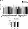
Compound #5 induces Gli1 expression. Female, 12‐week‐old C3H mice were treated with Cremophor or compound #5. Twenty‐four hours after drug treatment the small intestines and the proximal portion of the duodenum was harvested and subjected to qRT‐PCR analysis using a custom intestinal stem cell PCR primer array. Treatment with compound #5 significantly downregulated the expression of Cyr61, Axin2, Ctgf, Smoc2, Oct4, Yap1, Dach1, Msi1, and HeyL (A) but led to an upregulation of Gli1 (B; *, P < .05, Student's t test)
Figure 7
Figure 7
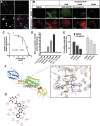
Compound #5 binds to Smoothened and activates Gli signaling. A, HEK293 cells were transiently transfected with an expression construct for mCherry‐tagged Smoothened protein. Smoothened‐mCherry overexpressing cells (red) were treated with BODIPY‐cyclopamine (green), fixed, counterstained with DAPI and imaged using confocal microscopy (×63 magnification, scale bar: 50 μM). The BODIPY signal colocalized with the Smoothened protein. B, HEK293 cells, overexpressing Smoothened‐mCherry were pretreated with BODIPY‐cyclopamine (green). Treatment with compound #5 led to a concentration‐dependent replacement of cyclopamine from its binding site. Images show clusters of HEK293 cells at ×4 magnification. Scale bar: 200 μM. C, Quantification of the replacement of BODIPY‐cyclopamine by compound #5 in mCherry‐tagged Smoothened protein overexpressing HEK293 using flow cytometry determined an IC50 of 88 μM ± 3.7 μM for compound #5 (three biologically independent repeats). D, Compound #5 caused a concentration‐dependent activation of a Gli signaling (*, P < .05 and **, P < .01, Student's t test). E, Compound #5 significantly attenuated the inhibitory effect of the Smoothened inhibitor Vismodegib in a budding enteroid assay. F, In silico docking results for compound #5 predicted binding to the transmembrane domain of Smoothened. G, LigPlot 2D presentation of the interactions of compound #5 with the transmembrane domain of Smoothened

Similar articles

Cited by

References

    1. Warren S, Bowers JZ. The acute radiation syndrome in man. Ann Intern Med. 1950;32:207‐216. - PubMed
    1. Weisdorf D, Chao N, Waselenko JK, et al. Acute radiation injury: contingency planning for triage, supportive care, and transplantation. Biol Blood Marrow Transplant. 2006;12:672‐682. - PubMed
    1. Withers HR, Elkind MM. Radiosensitivity and fractionation response of crypt cells of mouse jejunum. Radiat Res. 1969;38:598‐613. - PubMed
    1. Withers HR, Elkind MM. Microcolony survival assay for cells of mouse intestinal mucosa exposed to radiation. Int J Radiat Biol Relat Stud Phys Chem Med. 1970;17:261‐267. - PubMed
    1. Sato T, Vries RG, Snippert HJ, et al. Single Lgr5 stem cells build crypt‐villus structures in vitro without a mesenchymal niche. Nature. 2009;459:262‐265. - PubMed

Publication types