Review
. 2019 Sep;20(9):1368-1380.
doi: 10.3348/kjr.2019.0057.

A Stepwise Diagnostic Approach to Cystic Lung Diseases for Radiologists

Affiliations
Review

A Stepwise Diagnostic Approach to Cystic Lung Diseases for Radiologists

Kyu Chong Lee et al. Korean J Radiol. 2019 Sep.

Abstract

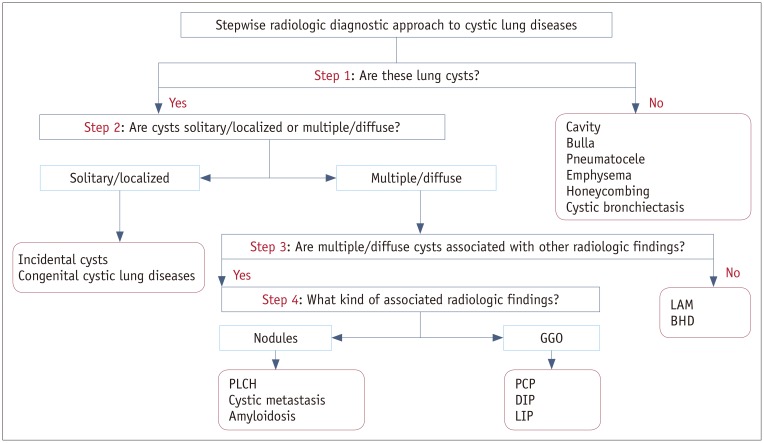
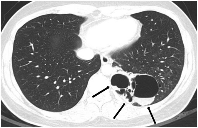
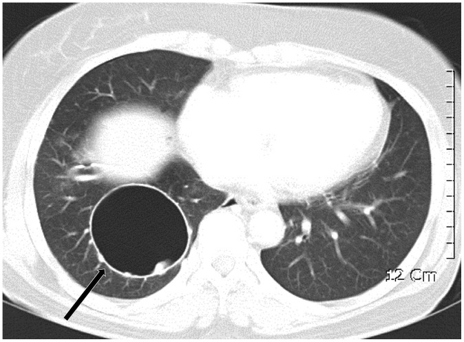
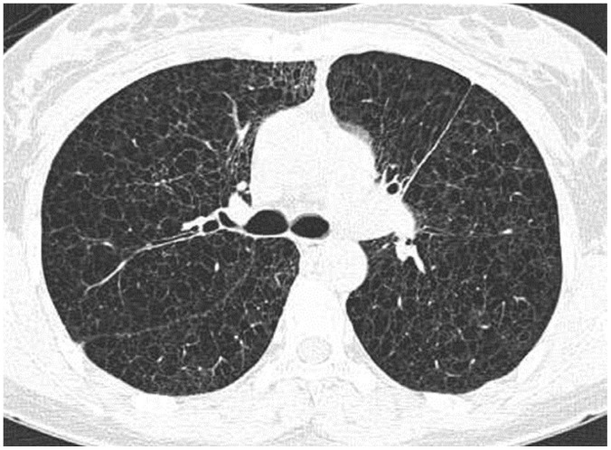
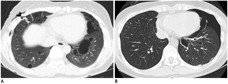
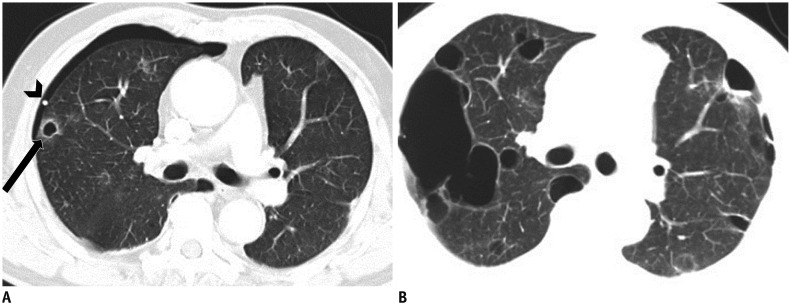
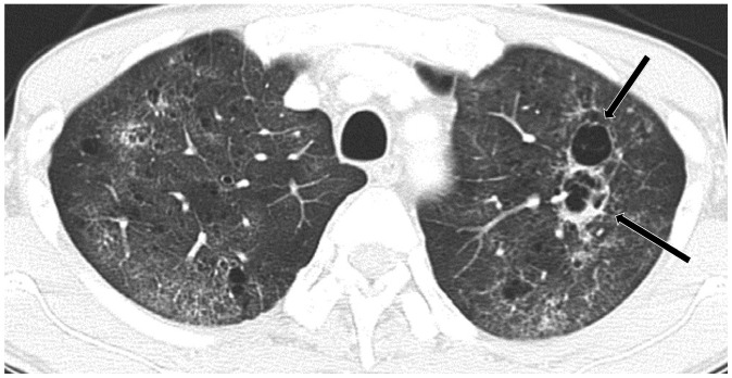
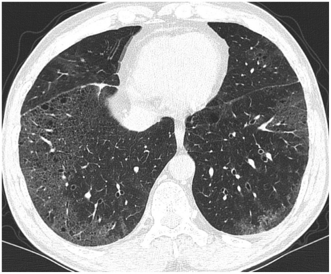
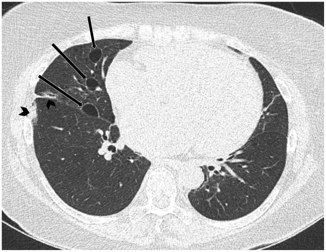
Lung cysts are commonly seen on computed tomography (CT), and cystic lung diseases show a wide disease spectrum. Thus, correct diagnosis of cystic lung diseases is a challenge for radiologists. As the first diagnostic step, cysts should be distinguished from cavities, bullae, pneumatocele, emphysema, honeycombing, and cystic bronchiectasis. Second, cysts can be categorized as single/localized versus multiple/diffuse. Solitary/localized cysts include incidental cysts and congenital cystic diseases. Multiple/diffuse cysts can be further categorized according to the presence or absence of associated radiologic findings. Multiple/diffuse cysts without associated findings include lymphangioleiomyomatosis and Birt-Hogg-Dubé syndrome. Multiple/diffuse cysts may be associated with ground-glass opacity or small nodules. Multiple/diffuse cysts with nodules include Langerhans cell histiocytosis, cystic metastasis, and amyloidosis. Multiple/diffuse cysts with ground-glass opacity include pneumocystis pneumonia, desquamative interstitial pneumonia, and lymphocytic interstitial pneumonia. This stepwise radiologic diagnostic approach can be helpful in reaching a correct diagnosis for various cystic lung diseases.

Keywords: Birt-Hogg-Dubé syndrome; Computed tomography; Histiocytosis; Langerhans cell; Lung diseases; Lymphangioleiomyomatosis.


Conflict of interest statement

The authors have no potential conflicts of interest to disclose.

Figures

Fig. 1
Fig. 1. Stepwise radiologic diagnostic approach to cystic lung diseases.
BHD = Birt-Hogg-Dubé syndrome, DIP = desquamative interstitial pneumonia, GGO = ground-glass opacity, LAM = lymphangioleiomyomatosis, LIP = lymphoid interstitial pneumonia, PCP = pneumocystis jirovecii pneumonia, PLCH = pulmonary Langerhans cell histiocytosis
Fig. 2
Fig. 2. Single or several air-filled lesions in localized area.
A. Cyst: round air-filled lesion with well-defined thin wall surrounded by normal lung (arrow). B. Cavity: air-filled lesion with thick wall within mass. C. Bulla: air-filled lesion, more than 1 cm in diameter, bounded by very thin imperceptible wall and associated with adjacent centrilobular emphysema. D. Pneumatocele: thin-walled, air-filled lesion (arrow) caused by pneumonia.
Fig. 3
Fig. 3. Multiple or numerous air-filled lesions distributed in both lungs.
A. Multiple and diffuse cysts in PLCH: multiple cysts of variable sizes and shapes with thin wall. B. Centrilobular emphysema: centrilobular lucencies without distinct walls, central dot within lucency represents branch of pulmonary artery. C. Honeycombing: multiple rows of air-filled spaces with thick wall clustered in subpleural region. D. Cystic bronchiectasis: tubular rather than spherical dimensions with branching pattern and associated bronchial wall thickening.
Fig. 4
Fig. 4. Incidental cyst in 73-year-old woman.
CT shows round 16-mm-sized thin-walled cyst (arrow) in right middle lobe.
Fig. 5
Fig. 5. Congenital pulmonary airway malformation in 18-year-old woman.
CT shows complex cystic mass (arrows) in left lower lobe, where largest cyst is larger than 2 cm. Lobectomy confirmed congenital pulmonary airway malformation.
Fig. 6
Fig. 6. Bronchogenic cyst in 55-year-old woman.
CT shows well-defined air-filled cyst (arrow) in right lower lobe, suggesting bronchogenic cyst. Surgical resection confirmed bronchogenic cyst.
Fig. 7
Fig. 7. Lymphangioleiomyomatosis in 28-year-old woman.
CT shows numerous cysts in both lungs. Cysts are round or ovoid and relatively uniform in size and shape. Cysts are diffusely distributed without zone predominance in both lungs. Lung biopsy was done through video-assisted thoracic surgery and lymphangioleiomyomatosis was confirmed histologically.
Fig. 8
Fig. 8. Birt-Hogg-Dubé syndrome with recurrent pneumothorax in 47-year-old woman (A) and 22-year-old woman (B) who were mother and daughter.
A. CT shows multiple, variable-sized, thin-walled cysts with lower subpleural lung predominance. Chest drainage tube (arrow head) is noted in right hemithorax for pneumothorax. B. CT shows multiple thin-walled cysts of various sizes and shapes in both lungs, with left lower lung dominance, and pneumothorax in left hemithorax.
Fig. 9
Fig. 9. PLCH in 22-year-old male smoker.
CT shows multiple, irregular, and round, thick- and thin-walled cysts with small irregular nodules (arrows) in both lungs, sparing lung bases and costophrenic angles (not shown), suggestive of PLCH, which was subsequently confirmed histologically.
Fig. 10
Fig. 10. Cystic metastasis from scalp angiosarcoma with recurrent pneumothorax in 71-year-old man.
A. CT shows multiple cystic or cavitary nodules (arrow) that were histologically confirmed as metastasis. Pneumothorax is noted with drainage tube (arrow head). B. CT performed after 2 months shows variable-sized thin-walled cystic lesions in both lungs that greatly increased in size and number as compared with previous CT scan (A).
Fig. 11
Fig. 11. Pneumocystis jirovecii pneumonia in 46-year-old HIV infected man with left pneumothorax.
CT in upper lung zone shows multiple irregular thick-walled and thin-walled cysts (arrows) on background of bilateral diffuse GGO. HIV= human immunodeficiency virus
Fig. 12
Fig. 12. Desquamative interstitial pneumonia in 52-year-old smoker.
CT shows GGO with tiny cysts in both lungs, mainly lower peripheral lungs, almost symmetrically distributed. Surgical lung biopsy confirmed desquamative interstitial pneumonia histologically.
Fig. 13
Fig. 13. Lymphoid interstitial pneumonia in 51-year-old woman with Sjögren's syndrome.
CT shows diffuse faint GGO and several cysts (arrows) in right lung. Cysts are thin-walled and distributed randomly. Previous surgical scars (arrowheads) are noted in subpleural region of right middle and lower lobes.

References

    1. Hansell DM, Bankier AA, MacMahon H, McLoud TC, Müller NL, Remy J. Fleischner Society: glossary of terms for thoracic imaging. Radiology. 2008;246:697–722. - PubMed
    1. Gupta N, Vassallo R, Wikenheiser-Brokamp KA, McCormack FX. Diffuse cystic lung disease. Part II. Am J Respir Crit Care Med. 2015;192:17–29. - PMC - PubMed
    1. Ryu JH, Swensen SJ. Cystic and cavitary lung diseases: focal and diffuse. Mayo Clin Proc. 2003;78:744–752. - PubMed
    1. Gafoor K, Patel S, Girvin F, Gupta N, Naidich D, Machnicki S, et al. Cavitary lung diseases: a clinical-radiologic algorithmic approach. Chest. 2018;153:1443–1465. - PubMed
    1. Parkar AP, Kandiah P. Differential diagnosis of cavitary lung lesions. J Belg Soc Radiol. 2016;100:100. - PMC - PubMed

MeSH terms

Supplementary concepts